Qaasid News
Download Our App
Latest News from Pakistan
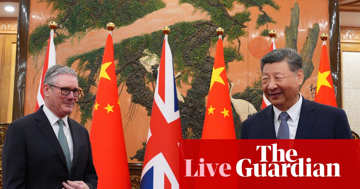
Starmer-Xi meeting live: UK prime minister says he wants ‘more sophisticated’ relationship with China | Keir Starmer
January 29, 2026
Reds Legends XV…Voting Race Tight in Key Positions
January 29, 2026
January 28, 2026 — Andrade Beats Swerve, Tommaso Ciampa Debuts, Champions Retain, More
January 29, 2026
At least 3 terrorists killed after security forces launch large-scale operation in Bannu’s Domel – Dawn
January 29, 2026
War, conflict and Roman sculptures: Bath exhibit shows different side of Don McCullin’s work | Don McCullin
January 29, 2026
The risky AI assistant clawing its way to viral fame – Morning Brew
January 29, 2026
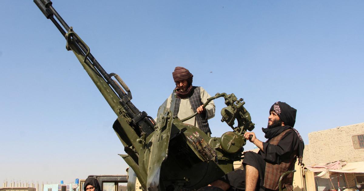
Afghanistan and Pakistan Square Off
January 29, 2026
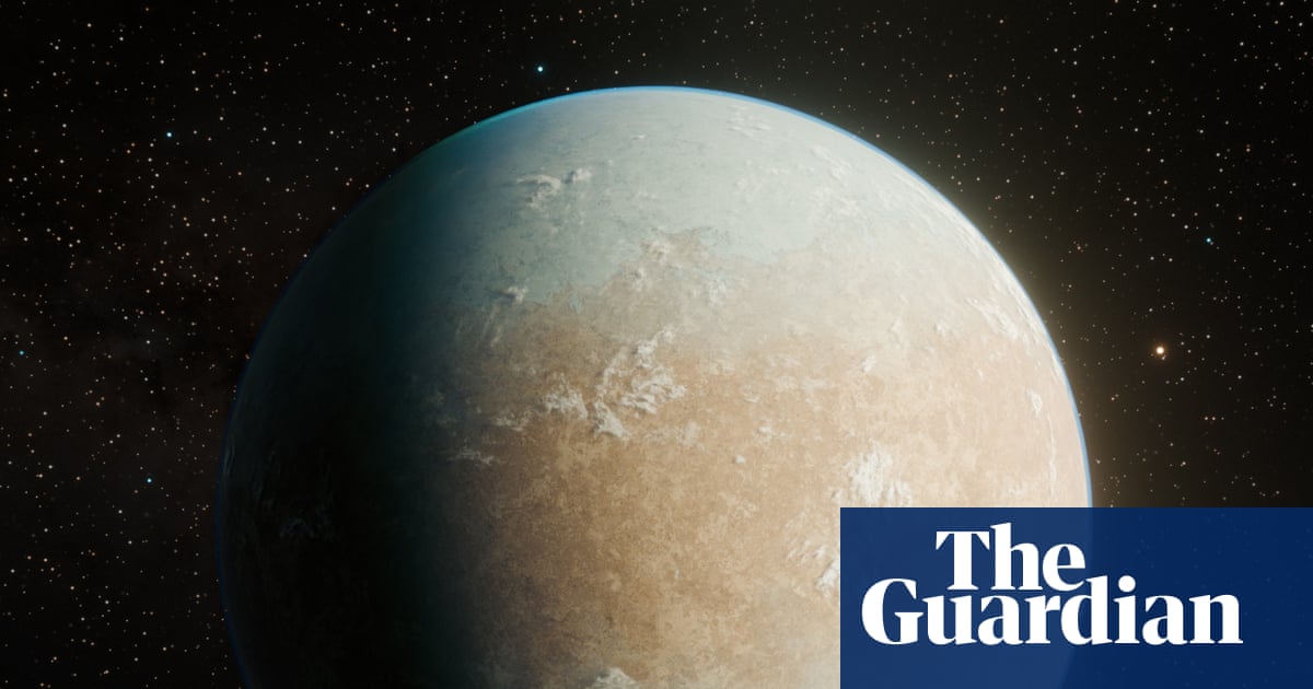
A potentially habitable new planet has been discovered 146 light-years away – but it may be -70C | Science
January 29, 2026
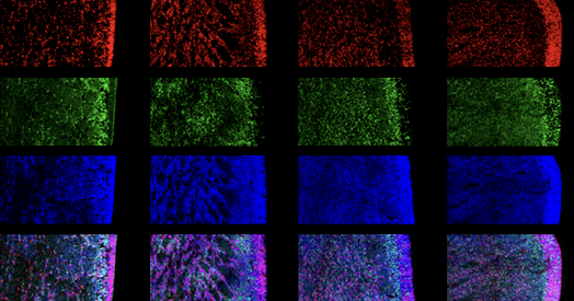
‘Tour de force’ study flags fount of interneurons in human brain
January 29, 2026
2025 Full year results | Givaudan
January 29, 2026