Qaasid News
Download Our App
Latest News from Pakistan
Feel like you gained 5 kilos right before your period? Nutritionist shares 5 nutrition hacks to manage PMS symptoms
December 11, 2025
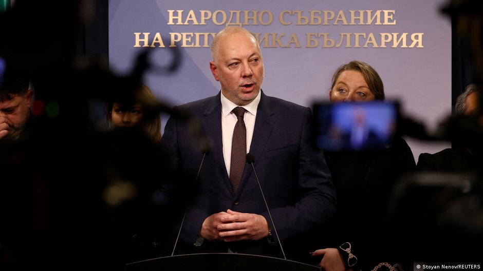
Bulgarian prime minister steps down after mass protests – DW – 12/11/2025
December 11, 2025
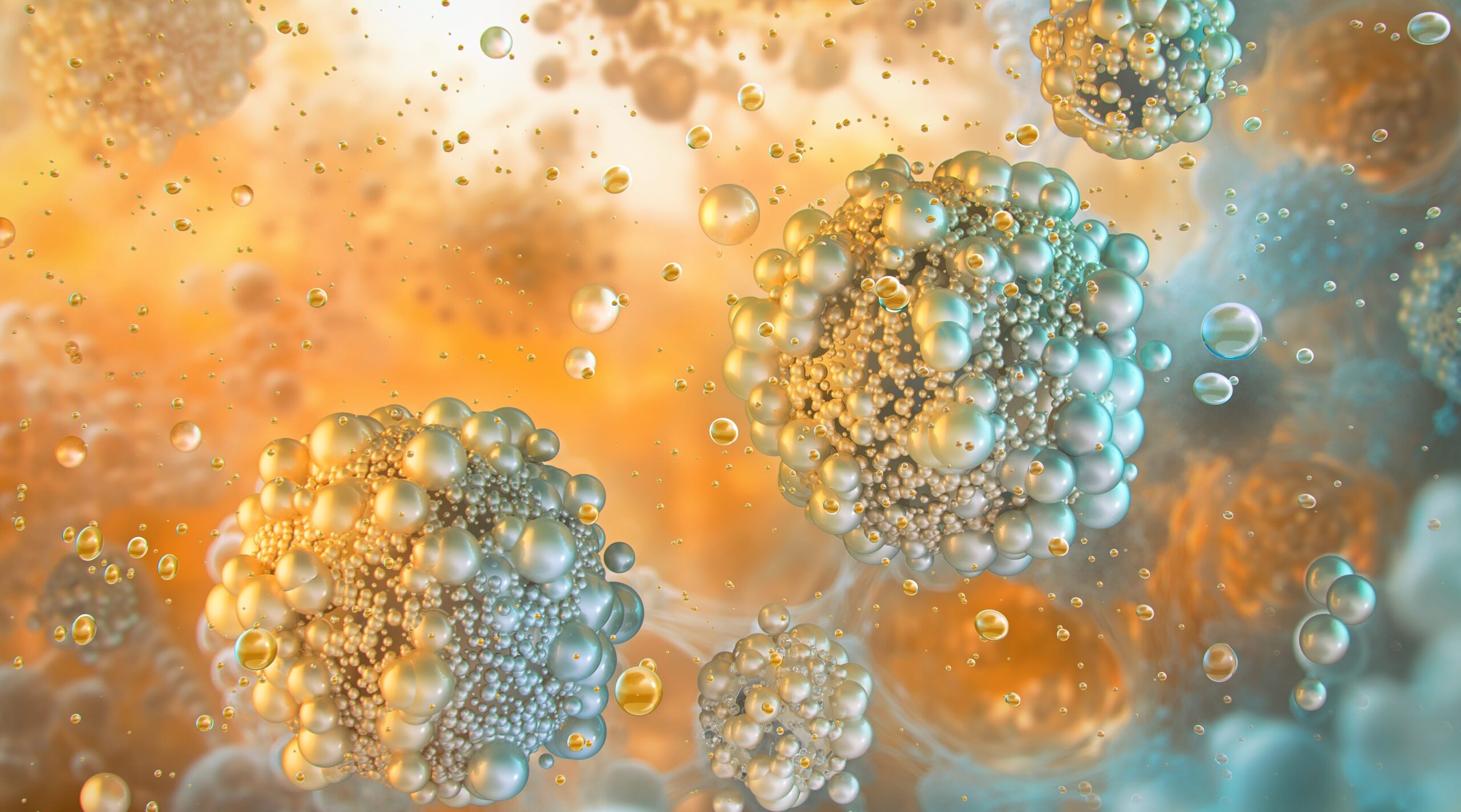
Platelet-inspired nanoparticles could improve treatment of inflammatory diseases
December 11, 2025
Bulgarian government resigns after mass anti-corruption protests | Bulgaria
December 11, 2025
Intel Core Ultra X9 388H Panther Lake CPU Gains Ground on AMD in Leaked Results – extremetech.com
December 11, 2025
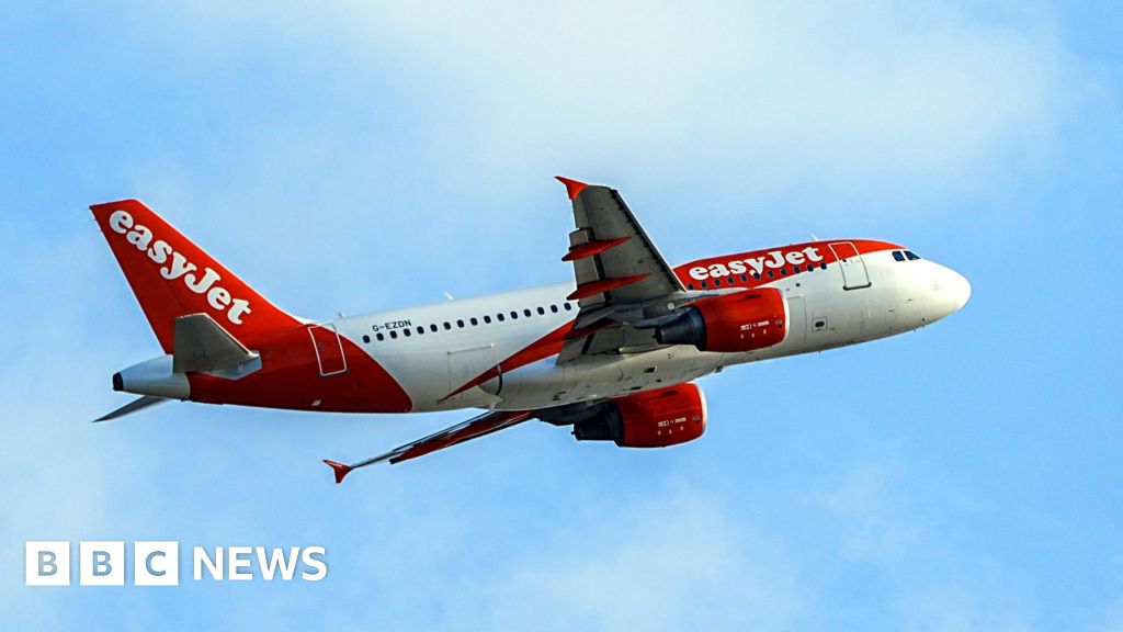
Guernsey States ‘in talks’ with Easyjet about island routes
December 11, 2025
Aleah Finnegan wins vault gold in Thailand
December 11, 2025
Mars CEO on How Business Can Be a Force for Good
December 11, 2025
Ferrero & Lopez win Coach of the Year in the 2025 ATP Awards – ATP Tour
December 11, 2025
Mars Completes Acquisition of Kellanova
December 11, 2025