Qaasid News
Download Our App
Latest News from Pakistan
The Under-the-Radar Brands Worn by Vogue’s Best Dressed List
December 11, 2025
New Photonic Techniques Aim to Break Three Longstanding Barriers to Quantum Scale
December 11, 2025
Bulgarian government collapses after weeks of mass protests – POLITICO
December 11, 2025
Nutritionist shares 5 essential nutrients parents need to add to children’s diet to protect them from seasonal flu
December 11, 2025
Apple’s New Patent Reveals a Modular Camera System With Interchangeable Lenses
December 11, 2025
EU watchdogs raid Temu’s Dublin HQ in foreign subsidy investigation | Business
December 11, 2025
Clinical characteristics of granular parakeratosis caused by benzalkon
December 11, 2025
Transcript of the Weekly Media Briefing by the Spokesperson on Thursday, 11 December 2025
December 11, 2025
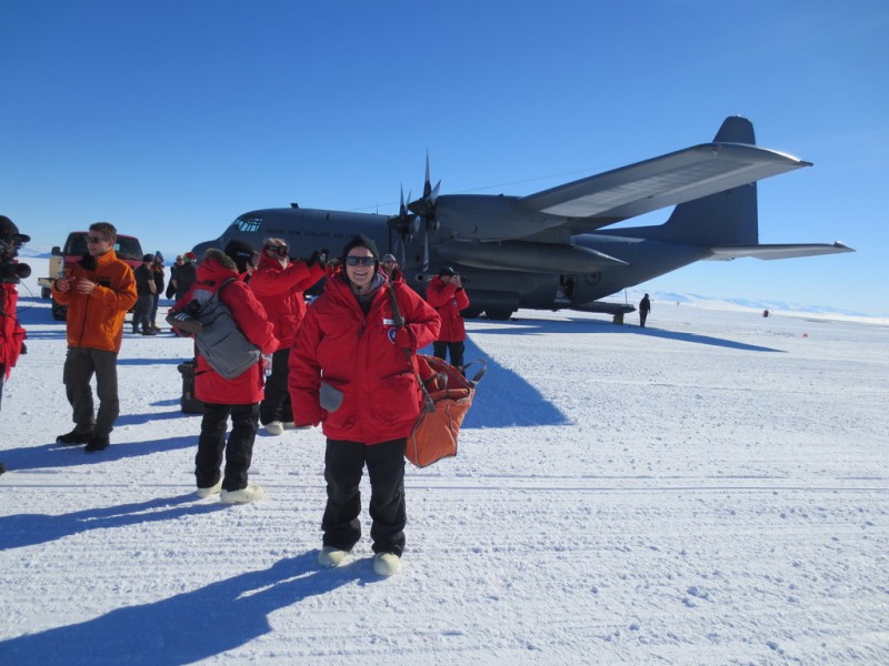
Scientist Hunts Ancient Climate Clues in Antarctic Ice
December 11, 2025
FCA’s Takeover of AML Supervision: Analysis for Law and Accountancy Firms : Clyde & Co
December 11, 2025