Qaasid News
Download Our App
Latest News from Pakistan
Australia leader defends social media ban as teens brag about staying online – Reuters
December 11, 2025
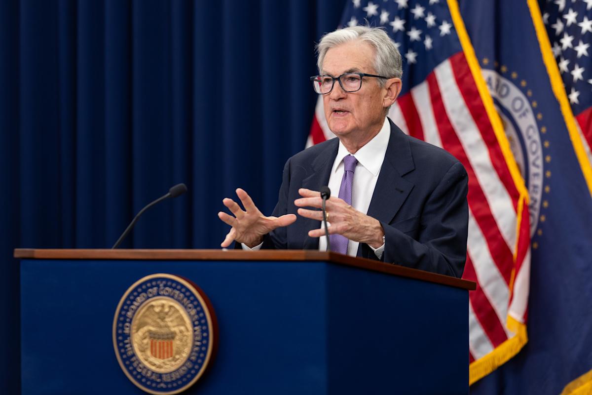
Federal Reserve cuts interest rates by 0.25%, Powell says there’s ‘no risk-free path’
December 11, 2025
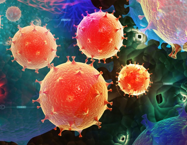
Hidden infections may play a key role in driving long COVID symptoms
December 11, 2025
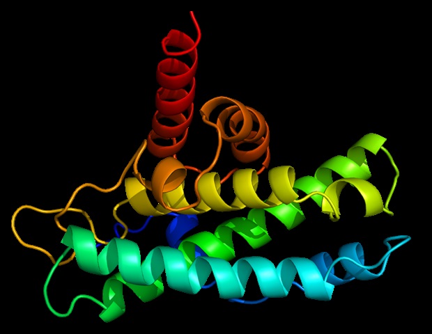
Study elucidates novel mitochondrial mechanisms underlying anti-aging and longevity
December 11, 2025
What’s likely to move the market in the next trading session
December 11, 2025
The Swiss city that lets you pay for most things with bitcoin
December 11, 2025
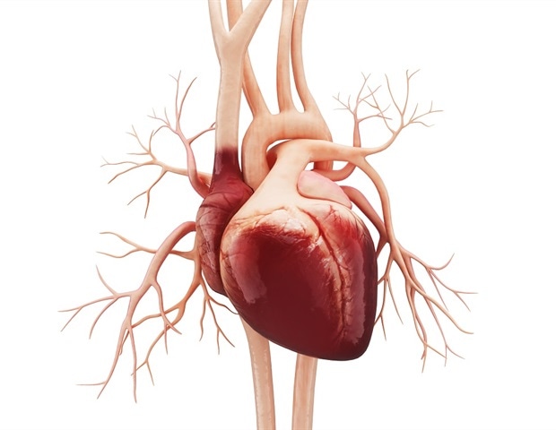
Family history of cardiometabolic disease linked to early heart damage in adolescents
December 11, 2025
Quantum Bumblebee Black Hole Radiative Properties And Particle Production For Spin-0, 1/2, 1, And 2 Fields
December 11, 2025
Apple CEO pushes for changes in US child online safety bill, citing privacy concerns – Reuters
December 11, 2025
A Deadly Coronavirus Resurfaces in France for First Time in 12 Years | The Transmission
December 11, 2025