Qaasid News
Download Our App
Latest News from Pakistan
Instagram now lets you see and control your algorithm
December 10, 2025
Astrin Launches Blood-Based Early Breast Cancer Detection Test – Inside Precision Medicine
December 10, 2025
BBVA Completes Share Buyback for Nearly €1 billion
December 10, 2025
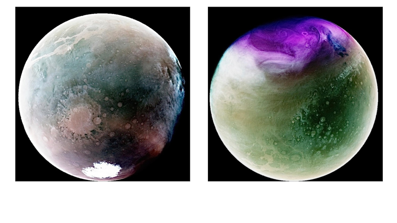
NASA loses contact with its Maven spacecraft orbiting Mars for the past decade
December 10, 2025
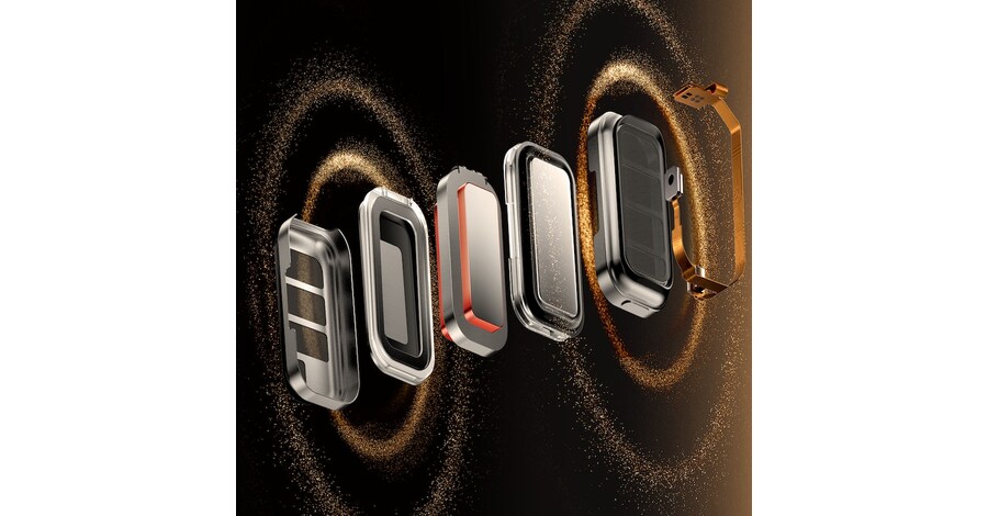
A Unified Technology System That Redefines Open-Ear Audio
December 10, 2025
Climate change is to blame for disappearing rains in the southwest
December 10, 2025
The 2026 CP+ Photo Show in Japan Will Be the Biggest Ever
December 10, 2025
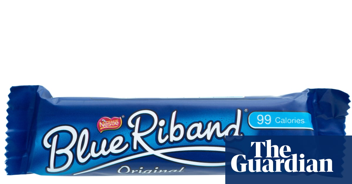
Toffee Crisp and Blue Riband no longer called ‘chocolate’ after recipe change | Chocolate
December 10, 2025
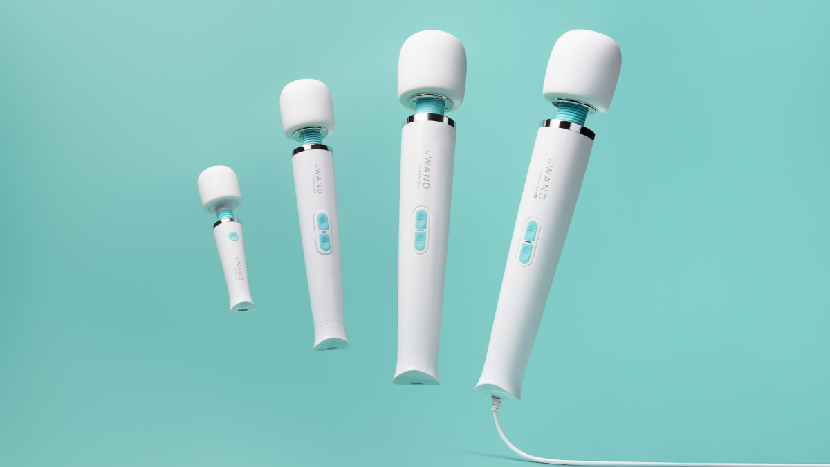
Le Wand launches Classique Collection: Get a free gift when you buy now
December 10, 2025
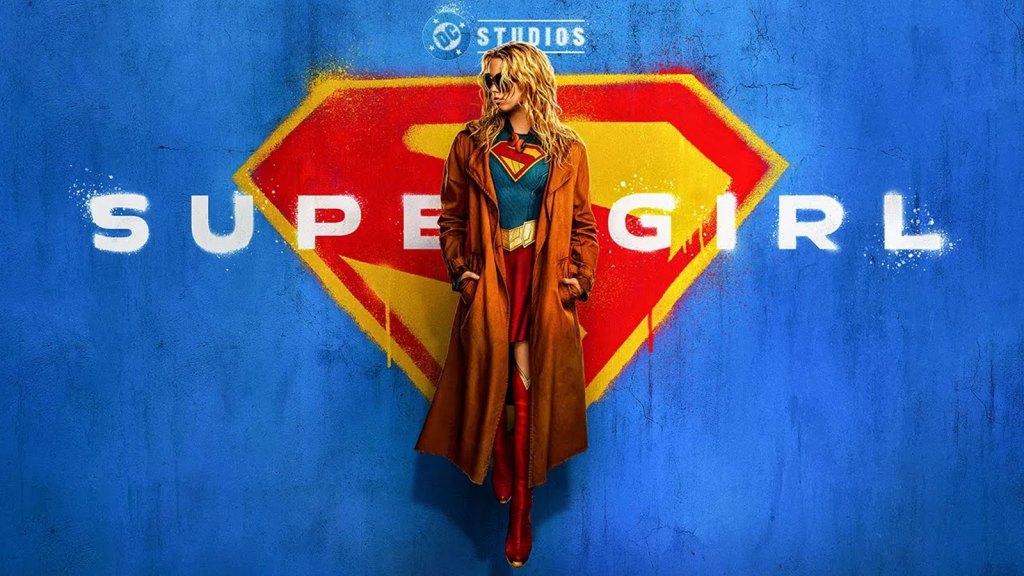
First Look ‘Supergirl’ Teaser Trailer For Milly Alcock-Starring DC Studios Film
December 10, 2025