Qaasid News
Download Our App
Latest News from Pakistan
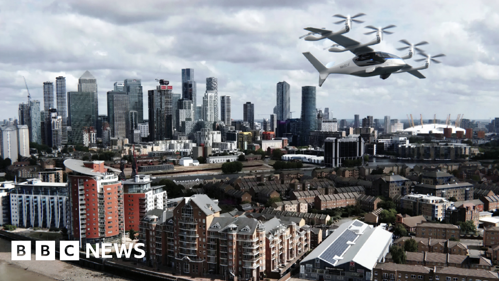
Flying taxis could be in London’s skies by 2028, developers say
December 10, 2025
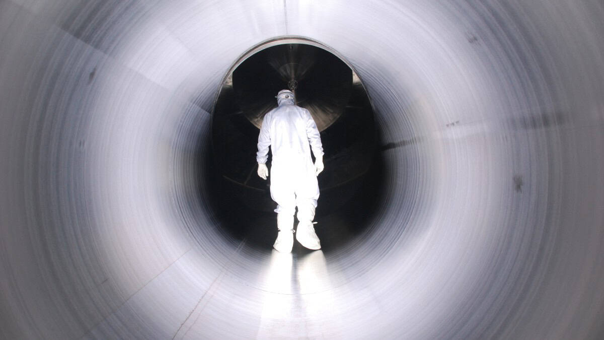
KATRIN and MicroBooNE come up empty handed – Physics World
December 10, 2025
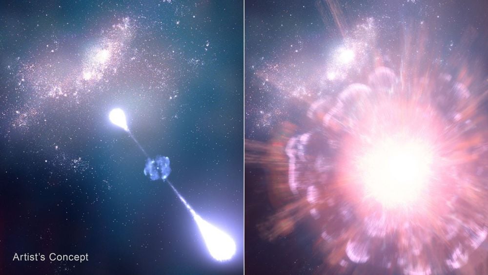
The JWST Just Identified A Supernova From Only 730 Million Years After The Big Bang
December 10, 2025
Tidal Love Numbers Reveal Nonzero Responses In Regular Black Holes, Offering A Window Into New Physics
December 10, 2025
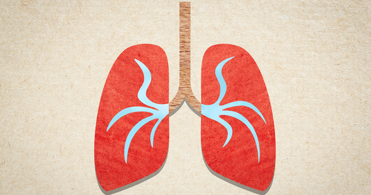
In pneumonia’s tug-of-war, lung microbiome could tip the balance
December 10, 2025
Meet Damhán Alla—the newly christened, spider-like feature on Jupiter's moon Europa – Phys.org
December 10, 2025
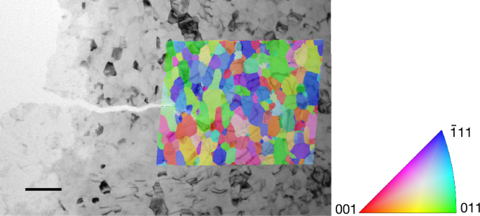
Quantifying grain boundary deformation mechanisms in small-grained metals
December 10, 2025
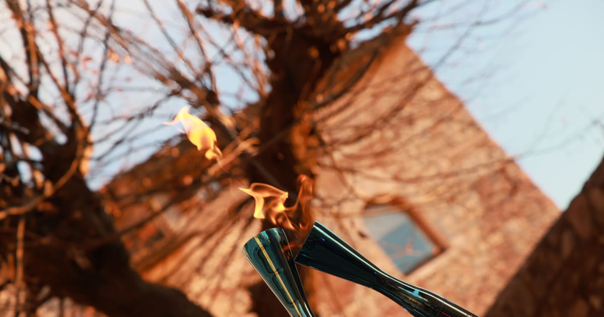
On Thursday the Milano Cortina 2026 Olympic Flame arrives in Firenze
December 10, 2025
First qualification systems for LA28 approved
December 10, 2025
A Multimodal AI Model May Improve Recurrence Risk Stratification in Early Breast Cancer
December 10, 2025