Qaasid News
Download Our App
Latest News from Pakistan
Malin Akerman Stars in May Cobb’s ‘All the Little Houses’ Audiobook
December 10, 2025
ALICE solves mystery of light-nuclei survival
December 10, 2025
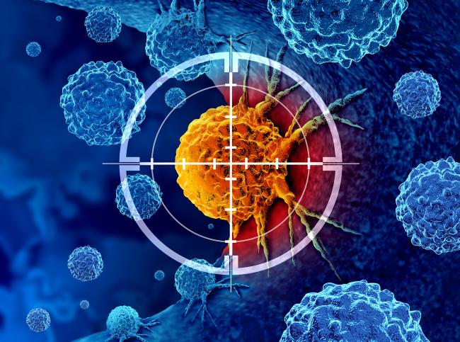
NIH-led study reveals role of mobile DNA elements in lung cancer progression
December 10, 2025
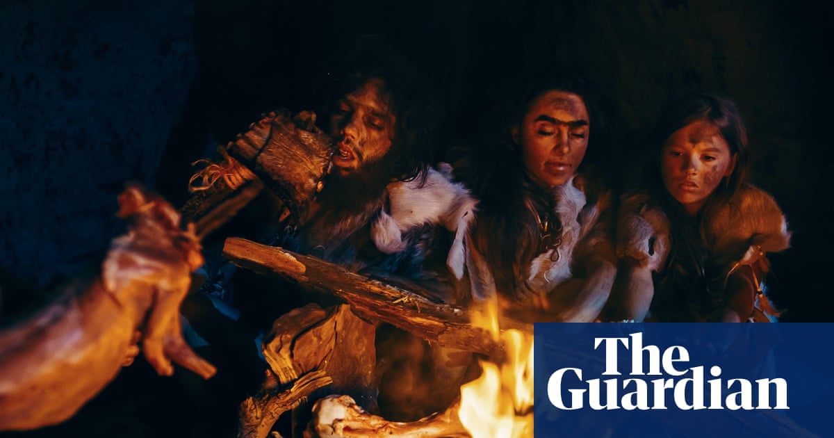
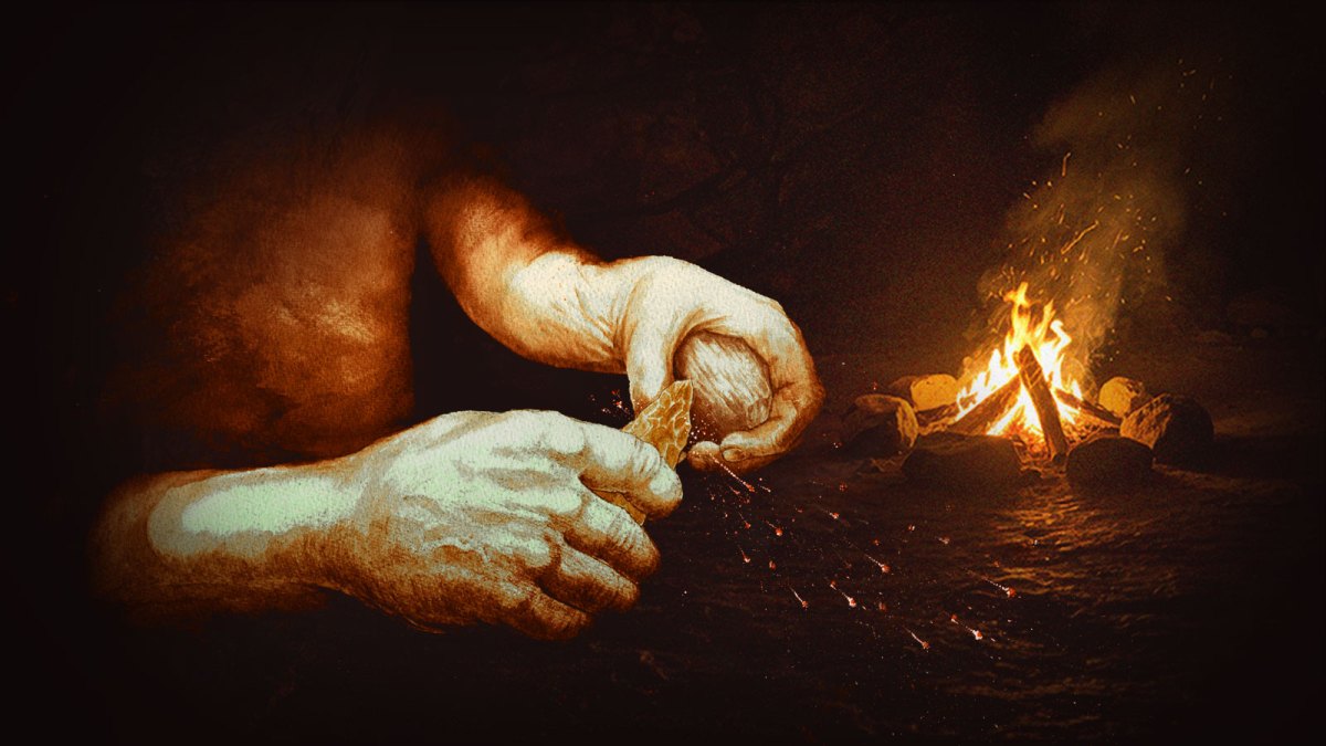
Man made fire 350,000 years earlier than previously thought, discovery in Suffolk suggests | Anthropology
December 10, 2025
First-trimester metformin use linked to pregnancy outcomes in PCOS patients
December 10, 2025
Spotlight On Belgrade As ‘The Librarians: The Next Chapter’ Location
December 10, 2025
Putin Pushes for Stronger Military and Energy Ties with Indonesia – Modern Diplomacy
December 10, 2025
British discovery rewrites history of when humans made fire – The Times
December 10, 2025
Blacklisted foreign organizations remain in Apple and Google app stores
December 10, 2025
World’s largest dinosaur track site discovered
December 10, 2025