Qaasid News
Download Our App
Latest News from Pakistan
Bestseller’s JJXX launches new looks featuring Spinnova fibre
January 27, 2026
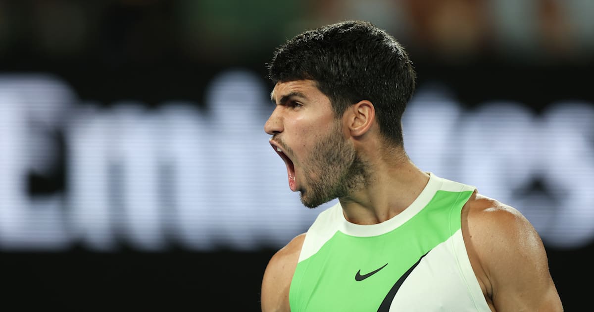
Carlos Alcaraz breaks Australian Open quarter-final duck by beating home hope Alex de Minaur
January 27, 2026
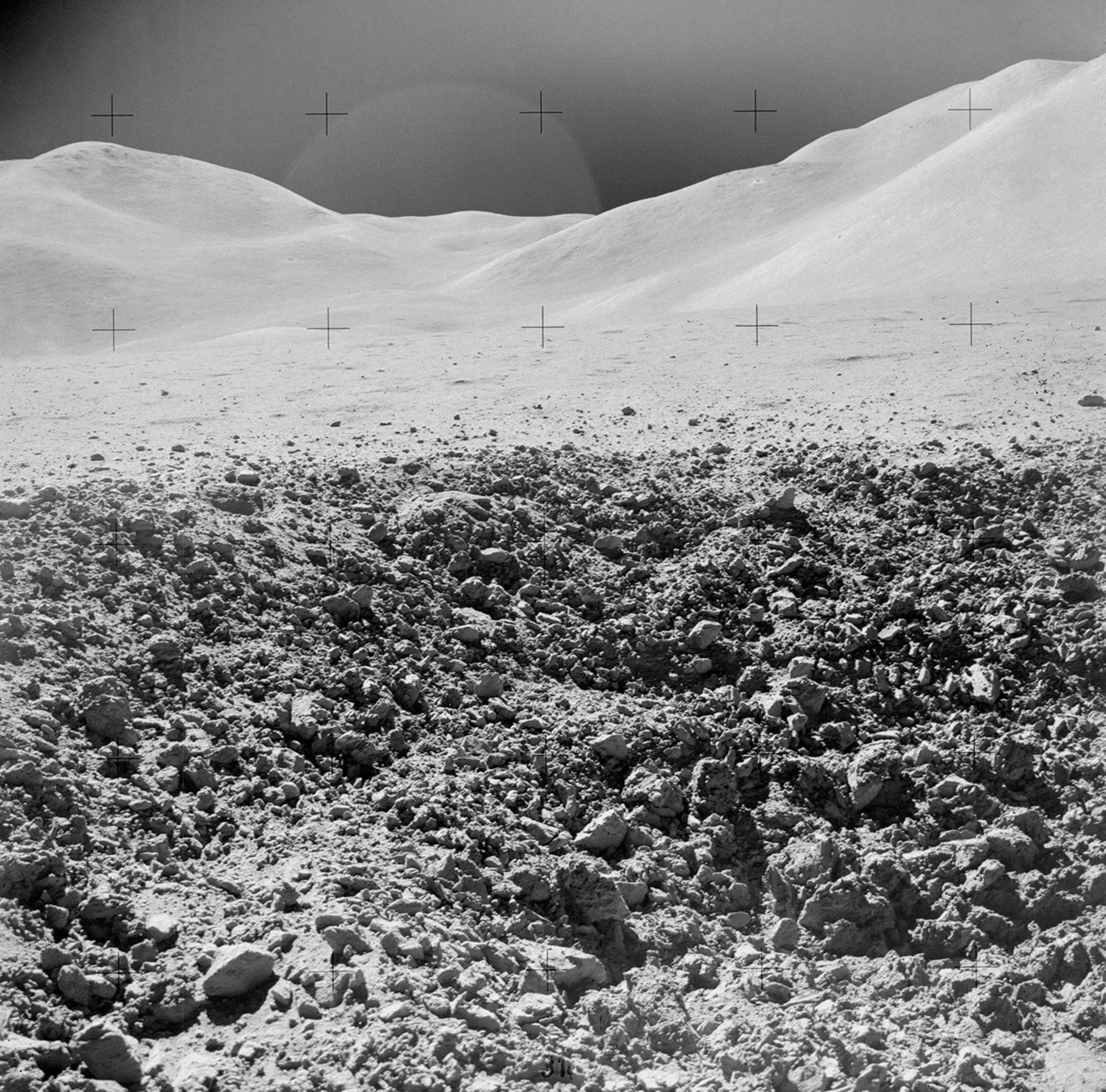
Moon samples put new constraints on meteorites as Earth’s water source
January 27, 2026
World Bank pushes Pakistan toward smarter revenue growth
January 27, 2026
Indonesia – Landslide, update (ADINet, media, BMKG) (ECHO Daily Flash of 27 January 2026) – ReliefWeb
January 27, 2026
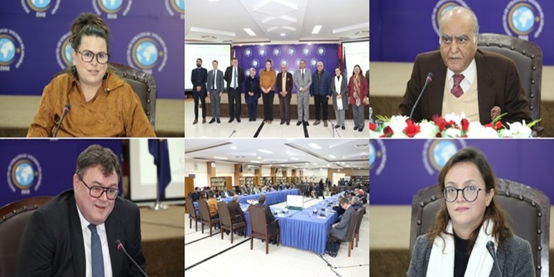
Press Release – ISSI Hosts Public Talk on “Europe, the Nordics, and the New Reality of Power Politics”
January 27, 2026
UK's Starmer heads to China to repair ties as he navigates tensions with US – Reuters
January 27, 2026
Karachi mall fire: Search operation ends, death toll rises to 73 – Press Trust of India
January 27, 2026
The West End’s Famous Free Late-Night Public Art Show Art After Dark Returns For 2026 Next Week
January 27, 2026
Teens charged over £11,000 phone thefts at Apple in Cambridge
January 27, 2026