Qaasid News
Download Our App
Latest News from Pakistan
Read An Excerpt Of Ye’s Letter That Blames A Brain Injury For His Antisemitism
January 26, 2026
L’anteprima della 51ma tappa del Viaggio della Fiamma Olimpica, da Dobbiaco a Bolzano
January 26, 2026
India, EU wrap up talks for landmark trade deal amid strained US ties – Reuters
January 26, 2026
KP CM pens letter to PM Shehbaz over Centre’s ‘persistent failure to release constitutionally guaranteed’ funds – Dawn
January 26, 2026
Champions League Matchday 8 possible line-ups and team news – UEFA.com
January 26, 2026
Apple Unveils Google Gemini-Powered Siri Next Month – 조선일보
January 26, 2026
UAE will not allow attacks on Iran from its soil: foreign ministry
January 26, 2026
Saudi Arabia wants ‘strong, positive’ relationship with UAE: FM – Dawn
January 26, 2026
PaleyFest L.A. to reunite ‘Charlie’s Angels’ actors for 50th anniversary
January 26, 2026
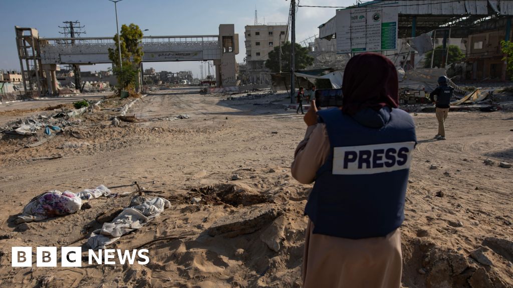
Israel court hears appeal against independent media access ban
January 26, 2026