Qaasid News
Download Our App
Latest News from Pakistan
Volkswagen, BMW and Mercedes cars could get cheaper as India plans EU tariff cuts
January 26, 2026
‘For the authoritarian, culture is the enemy’: Salman Rushdie talks recovery and resilience at Sundance | Sundance 2026
January 26, 2026
Solar Orbiter spots magnetic avalanches driving major solar flare
January 26, 2026
Metal rich winds detected in giant dusty cloud around distant star
January 26, 2026
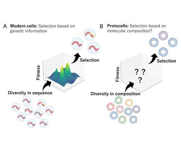
Icy cycles may have driven early protocell evolution
January 26, 2026
Seismic networks offer new way to track space junk reentering atmosphere
January 26, 2026
Private jet crashes in flames at Maine airport with eight aboard – Reuters
January 26, 2026
Amanat Holdings PJSC And 2 Other Undiscovered Gems In The Middle East
January 26, 2026
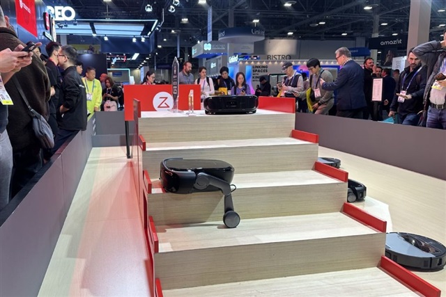
Roborock unveils stair-climbing vacuum as Chinese rivals heats up – digitimes
January 26, 2026
Who Is Imaan Mazari, Pak Lawyer Jailed For Anti-State Social Media Posts
January 26, 2026