Qaasid News
Download Our App
Latest News from Pakistan
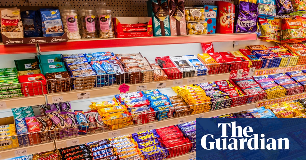
Almost a quarter of UK GPs are seeing obese children aged four and under | Obesity
January 25, 2026
‘I Want Your Sex’ Writer Karley Sciortino Reveals Inspiration Behind the Raunchy Sundance Film
January 25, 2026
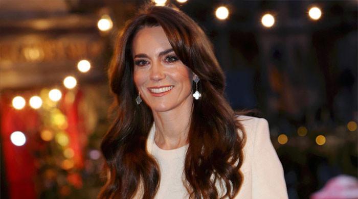
Princess Kate makes Burns Night a viral love letter to Scotland
January 25, 2026
Fujifilm Overtakes Canon in Amazon’s Mirrorless Best Sellers
January 25, 2026
The looming risk of food shortages and anarchy in the UK | Farming
January 25, 2026
Actors Hudson Williams and Connor Storrie light up Milano Cortina 2026 torch relay with pure joy
January 25, 2026
Brentford head coach Keith Andrews provides Kristoffer Ajer and Mikkel Damsgaard injury update
January 25, 2026
I tested a solid-state portable battery for a week – now lithium-ion feels old school
January 25, 2026
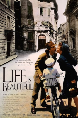
Life is Beautiful at Farm Street Film Club
January 25, 2026
Highlights from How I Treat Cervical Cancer in 2026
January 25, 2026