Qaasid News
Download Our App
Latest News from Pakistan
Justice Rozi, Arshad sworn in as Judges of FCC – RADIO PAKISTAN
November 17, 2025
Quinn Drops 30 as Cardinals Defeat Emerson, Winning Courtyard by Marriott Classic Title
November 17, 2025
Remnant cholesterol is associated with blood pressure control
November 17, 2025
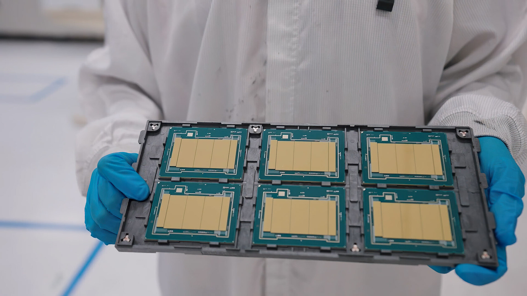
Intel “Granite Rapids-WS” Workstation Xeon SKUs Appear With Up to 336 MB of Cache
November 17, 2025
Samsung Galaxy S26 Ultra may skip the RAM boost you want
November 17, 2025
Bangladesh beefs up security ahead of verdict against ousted PM Sheikh Hasina – The Washington Post
November 17, 2025
Geely Automobile Net Profit Rises Sharply on Robust Sales
November 17, 2025
Two more judges take oath as Constitutional Court begins work
November 17, 2025
Efficacy analysis of a 12-cytokine panel for the diagnosis of Kawasaki
November 17, 2025
DPM to lead Pakistan delegation at SCO meeting in Moscow today – RADIO PAKISTAN
November 17, 2025