Qaasid News
Download Our App
Latest News from Pakistan
First Thing: Trump declaration of Greenland deal framework met with scepticism | US news
January 22, 2026
The Scoop with Nordstrom’s Jian DeLeon
January 22, 2026
Winter chills hit Karachi as rain, gusty winds signal three-day cold spell
January 22, 2026
GBH Daily: How to recognize AI videos
January 22, 2026
Israel bombs Lebanon-Syria border, 2 killed
January 22, 2026
Rapid rollback of Kurdish-led forces reshapes Sharaa’s Syria
January 22, 2026
How Ready to Advise rebuilt team confidence at Papin CPA
January 22, 2026
EU researchers inch closer to a viable quantum internet
January 22, 2026
Douglas, Martinez & Beddall in for Munster match
January 22, 2026
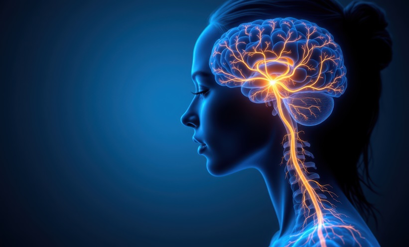
Vagus Nerve Stimulation In Resistant Depression
January 22, 2026