Qaasid News
Download Our App
Latest News from Pakistan
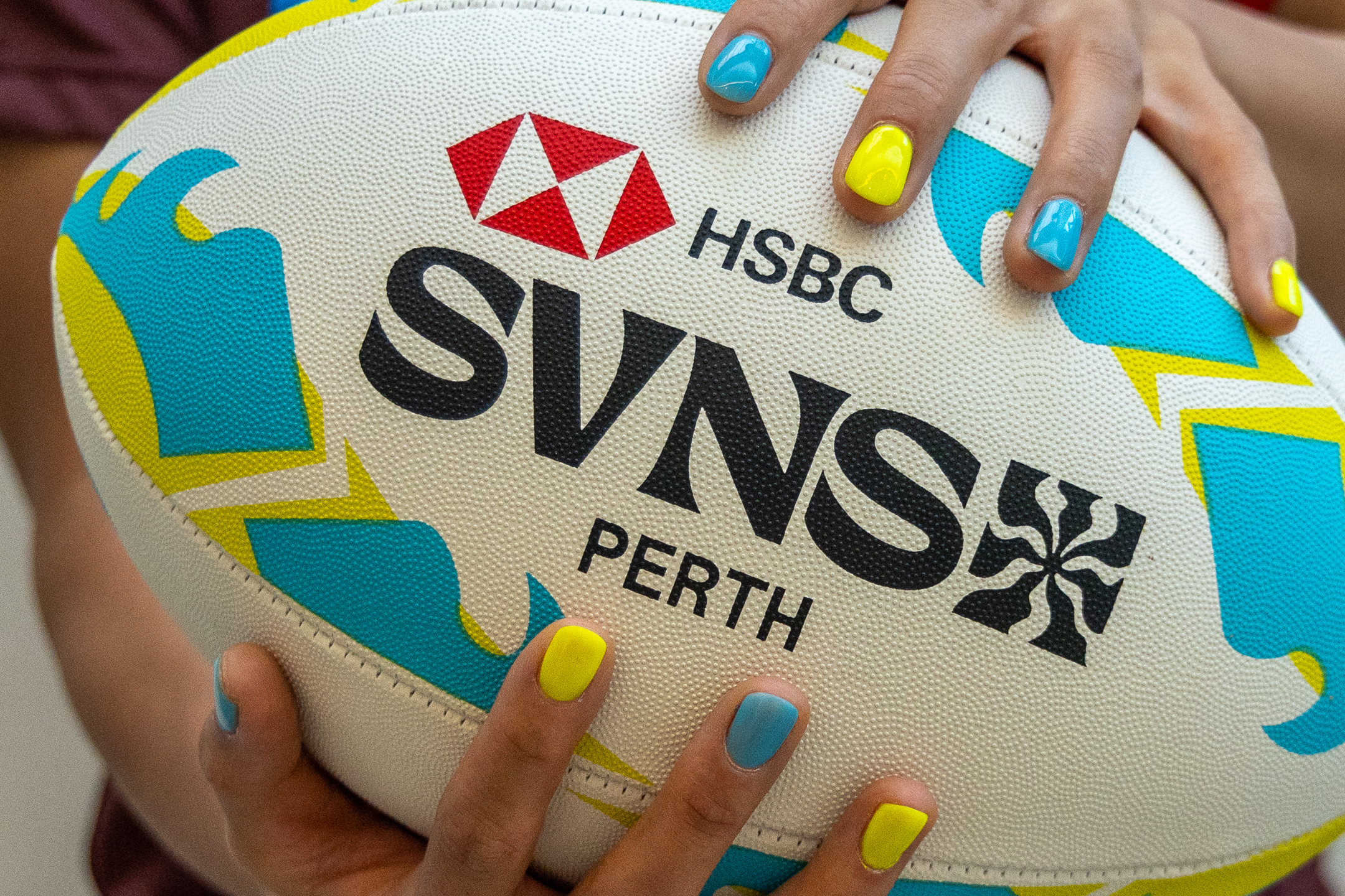
HSBC SVNS introduces Hard as Nails ahead of Perth weekend
February 5, 2026
Ukraine war briefing: Zelenskyy voices hope for new prisoner exchange as talks continue | Russia
February 5, 2026
The Strad News – Hong Kong concertmaster takes up guest role in China
February 5, 2026
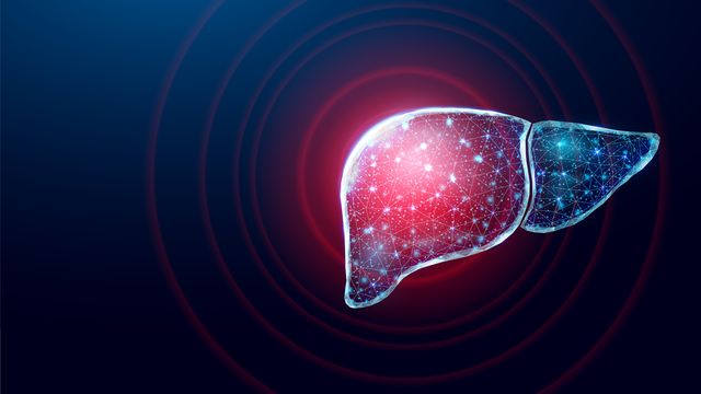
Liver Model Advances Drug Discovery for Fatty Liver Disease
February 5, 2026
Top health innovation role for Gates Cambridge Scholar
February 5, 2026
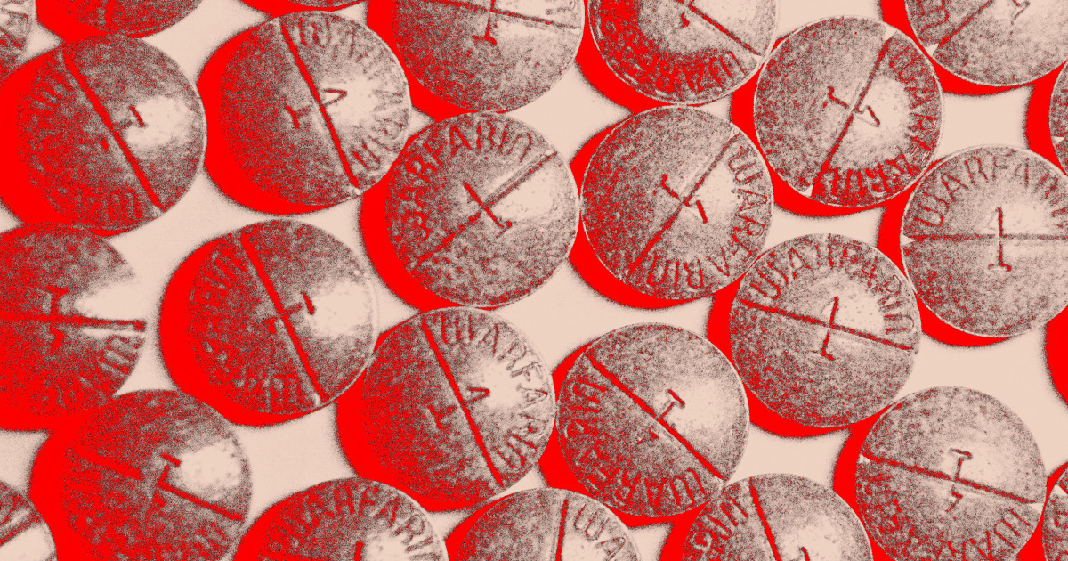
Blood thinners are a leading cause of drug-related harm. Can the risk be lowered?
February 5, 2026
Northern Lights Alert: 11 States May See Aurora Thursday – Forbes
February 5, 2026
What’s new to streaming this week? (Feb, 6, 2026)
February 5, 2026
Kashmir Solidarity Day: PID organizes roundtable discussion in Lahore – RADIO PAKISTAN
February 5, 2026
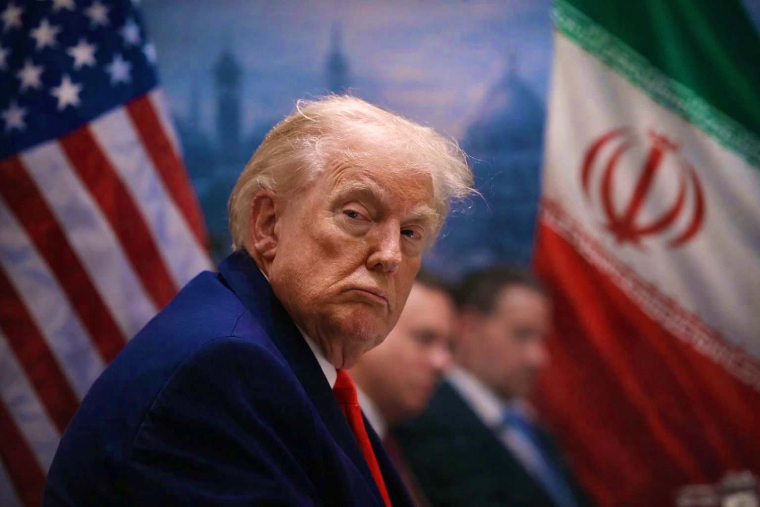
US Compellence Against Iran and the Limits of Pressure
February 5, 2026