Qaasid News
Download Our App
Latest News from Pakistan
Europeans prepare military exercises in Greenland, Trump’s ambitions undeterred – Dawn
January 15, 2026
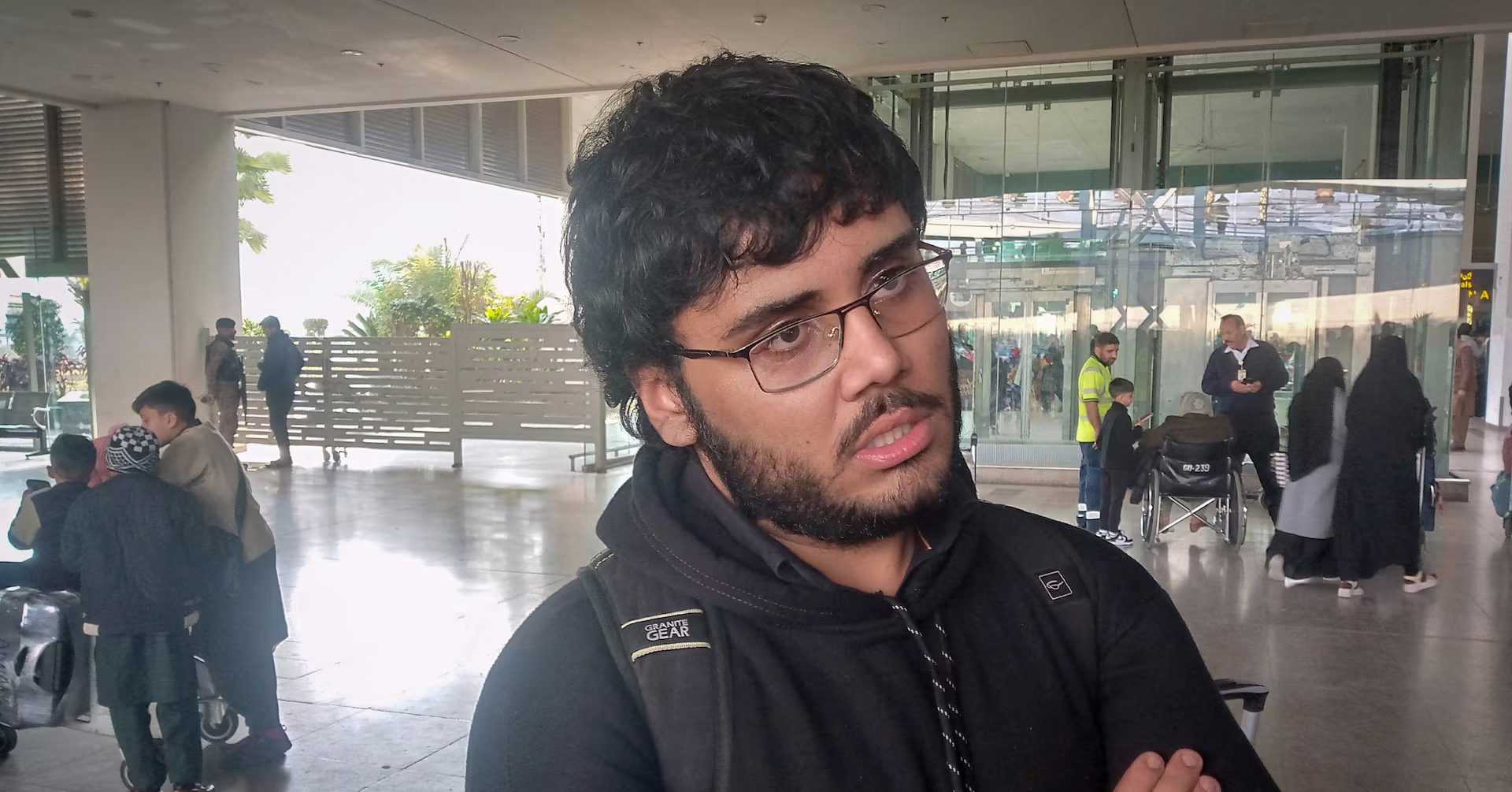
Back from Iran, Pakistani students say they heard gunshots while confined to campus
January 15, 2026
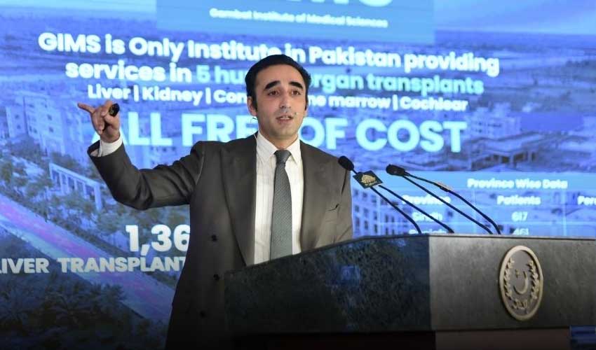
Bilawal says Sindh government works effectively across all sectors
January 15, 2026
Key Non-Polar Destinations Across the Moon to Address Decadal-level Science Objectives with Human Explorers
January 15, 2026
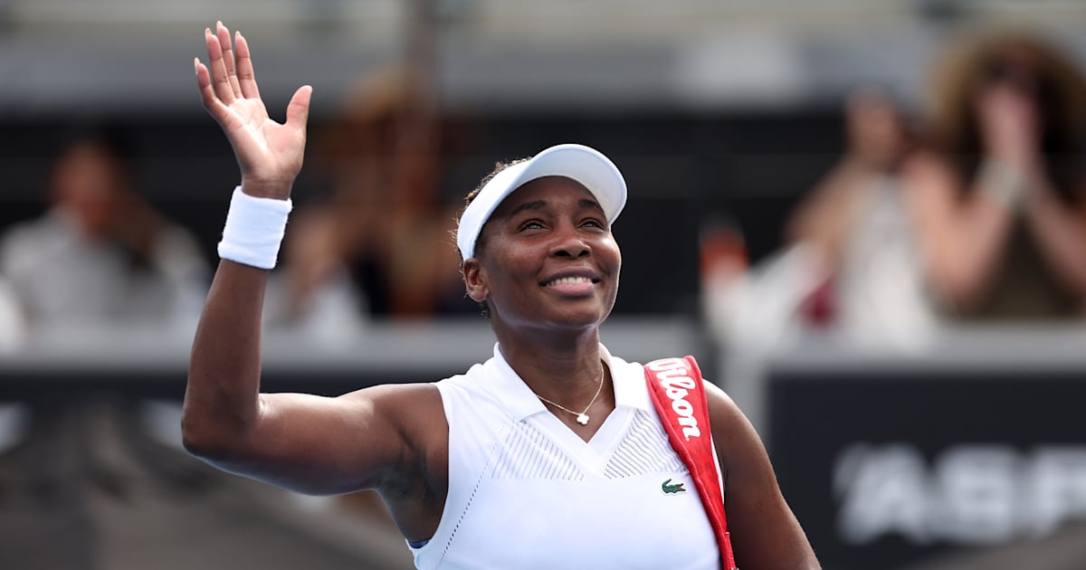
How to watch Venus Williams at the Australian Open 2026 – full schedule
January 15, 2026
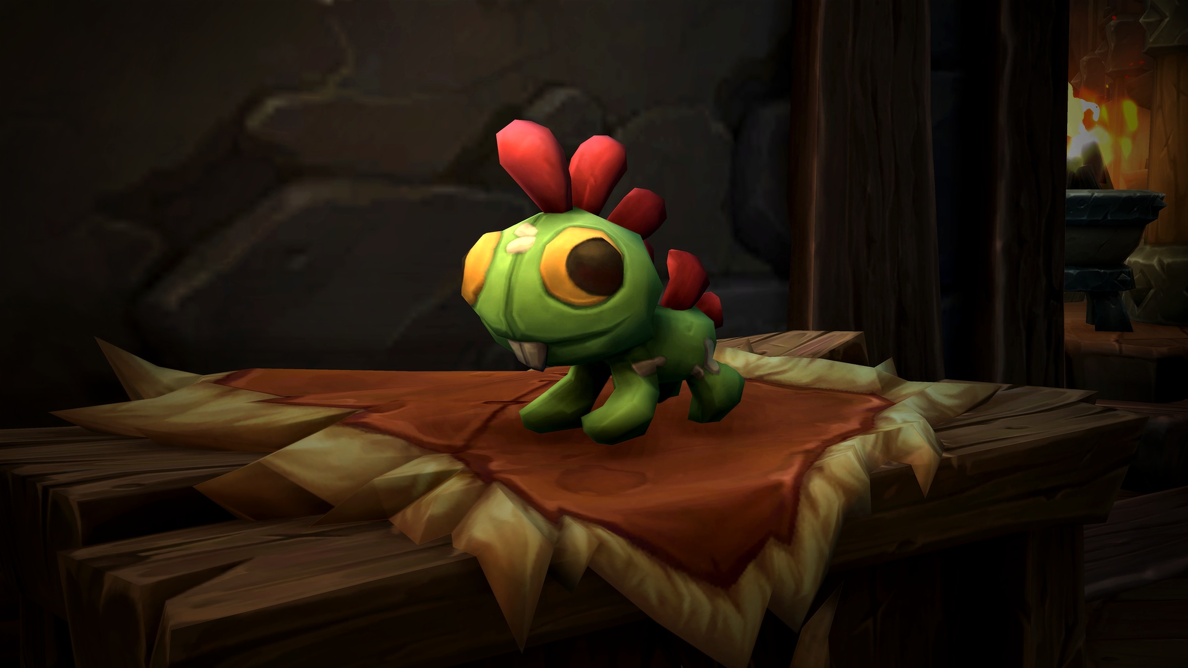
Get the Cuddly Green Grrgle Housing Decor Item January 20! — World of Warcraft — Blizzard News
January 15, 2026
Gulf states and Turkey urged Trump not to launch strikes against Iran | Iran
January 15, 2026
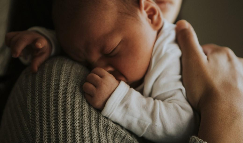
Finding moms’ vaccine-induced whooping cough antibodies in babies’ noses highlights benefits of indirect immunization
January 15, 2026
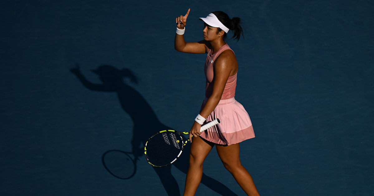
How to watch Alexandra Eala at the Australian Open 2026 – full schedule
January 15, 2026
Harry Styles announces fourth solo album, Kiss All the Time. Disco, Occasionally | Harry Styles
January 15, 2026