Qaasid News
Download Our App
Latest News from Pakistan
Astronauts set to leave ISS in first-ever medical evacuation – France 24
January 14, 2026
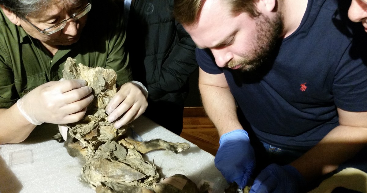
In the stomach of a mummified wolf pup, scientists find DNA from a woolly rhino
January 14, 2026
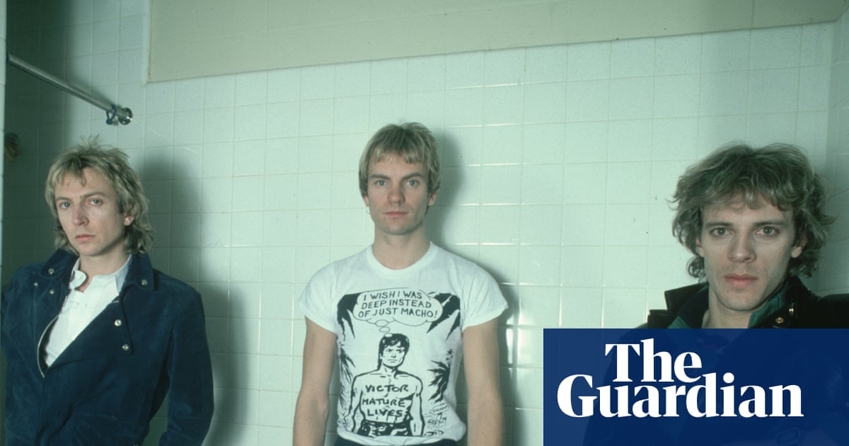
Can’t stand losing out: battle over the Police’s royalties reaches high court | UK news
January 14, 2026
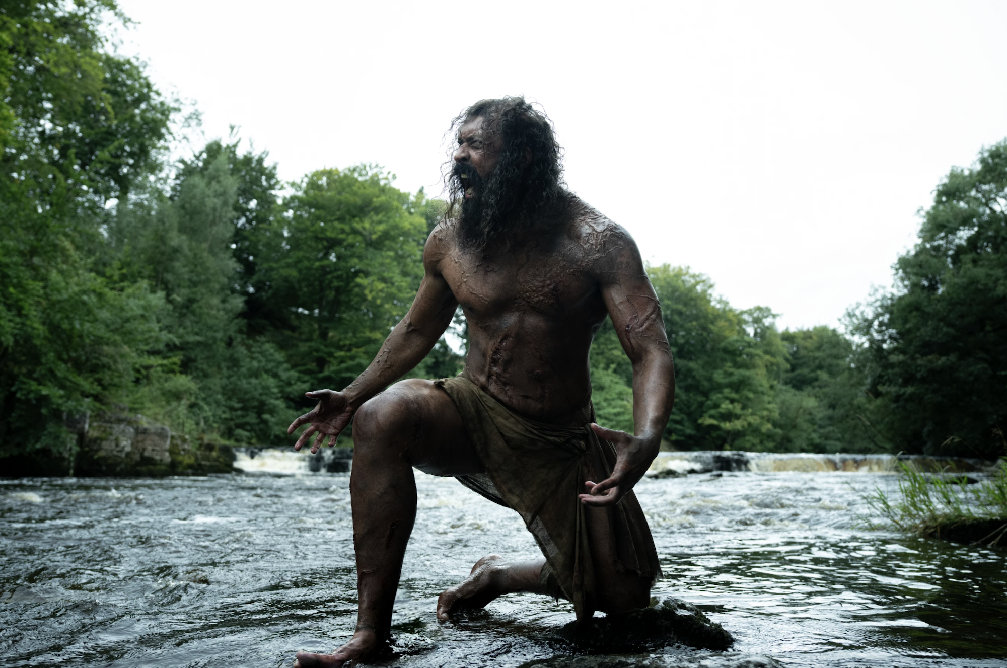
Movie Review: ‘28 Years Later: The Bone Temple’
January 14, 2026
How to watch Eileen Gu compete at Laax Open slopestyle World Cup ahead of Milano Cortina 2026
January 14, 2026
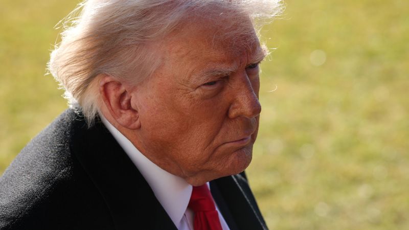
Live updates: Vance and Rubio to hold meeting on Greenland as Trump faces Venezuela war powers vote
January 14, 2026
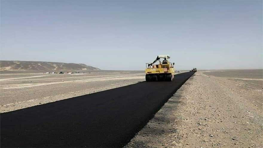
Work on Karachi–Chaman highway begins: PM
January 14, 2026
Stock market today: Live updates
January 14, 2026
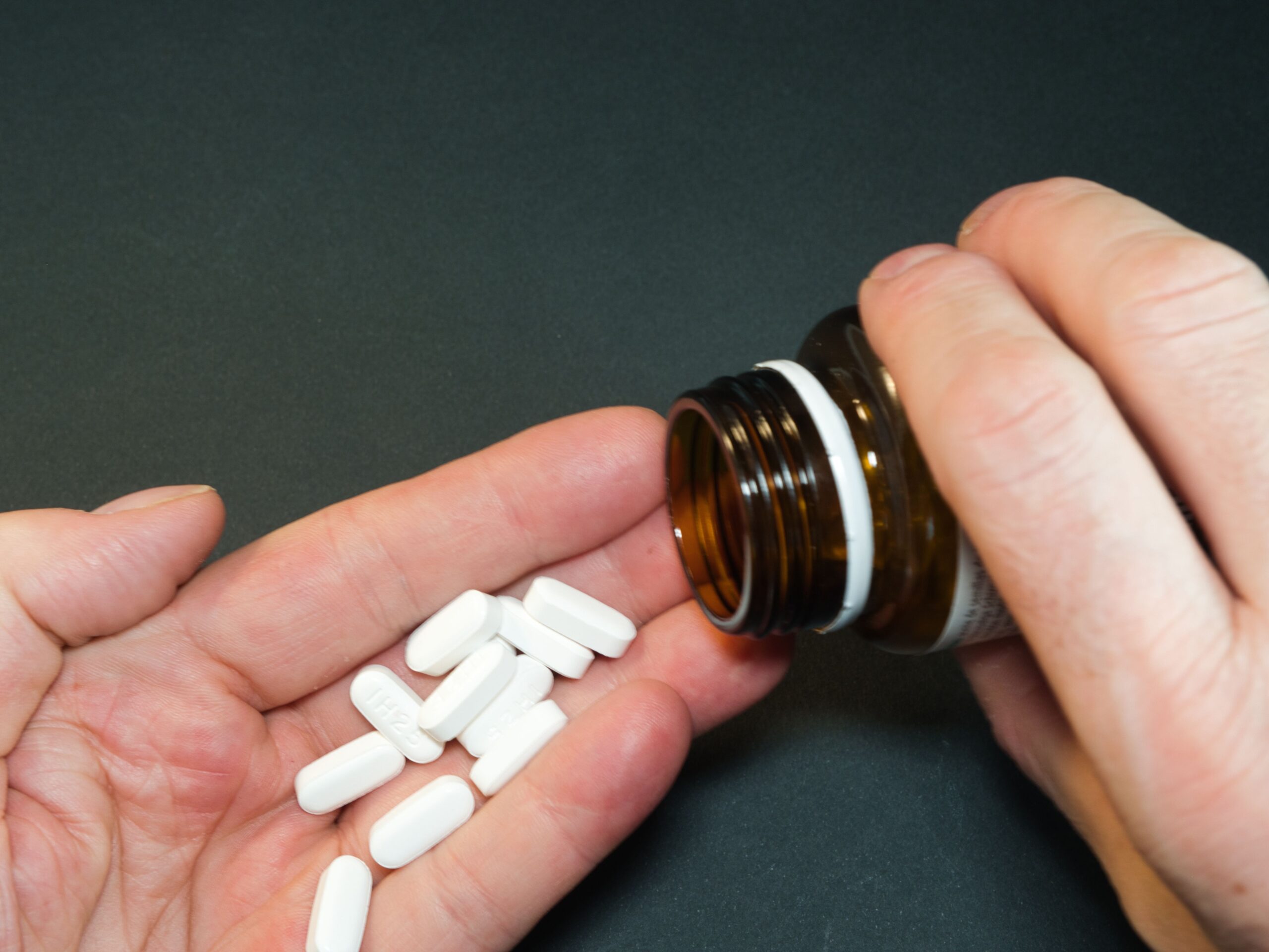
Strong Pharmacist Engagement Helps Patients Effectively Use OTC Products
January 14, 2026
California investigates Grok over AI deepfakes
January 14, 2026