Qaasid News
Download Our App
Latest News from Pakistan
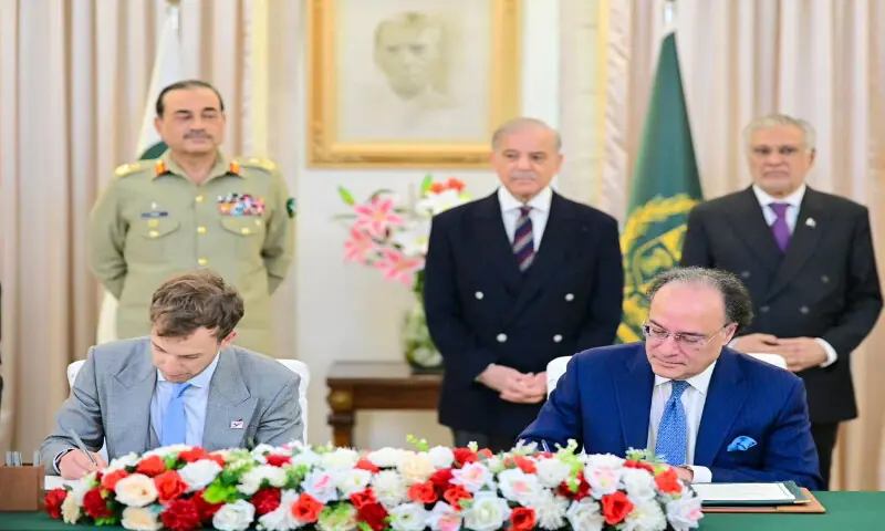
Pakistan signs deal with affiliate of World Liberty Financial on stablecoin – Pakistan
January 14, 2026
Ellie White: It’s important to start 2026 strong
January 14, 2026
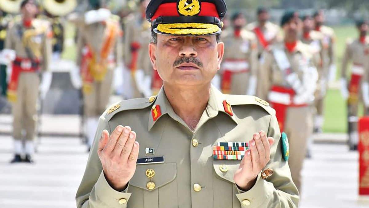
Panic in Pakistan as Trump backs regime change in Iran, Asim Munir holds emergency meeting – Firstpost
January 14, 2026
‘It Felt Like I Was Trespassing on the Privilege of Adventure Travel’
January 14, 2026
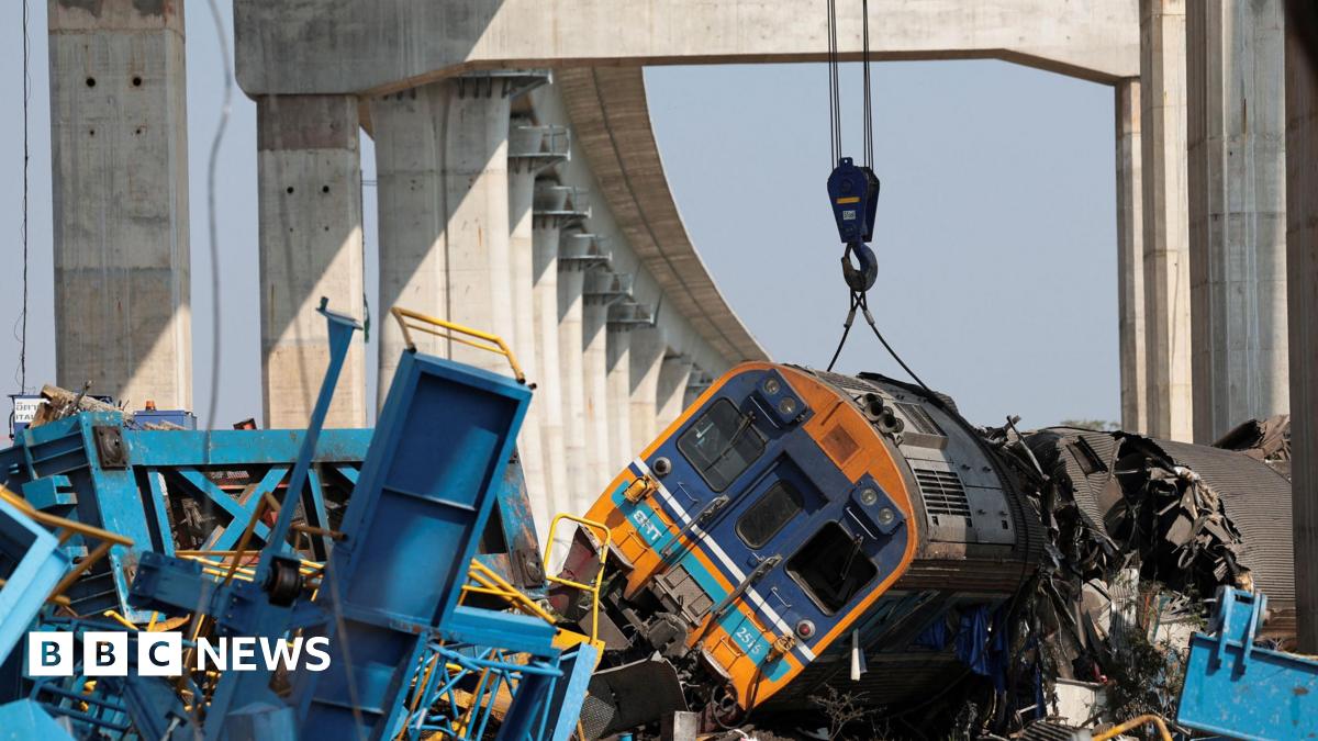
At least 22 dead as crane collapses onto train in Thailand – BBC
January 14, 2026
BRITs Week 2026 Lineup Announced – The BRIT Awards
January 14, 2026
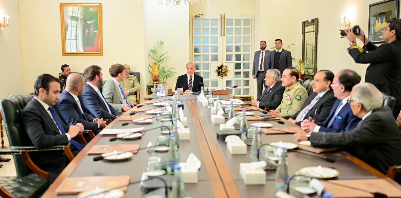
Rapid growth of digital payments, financial innovation, essential parts of Pakistan’s expanding digital economy: PM
January 14, 2026
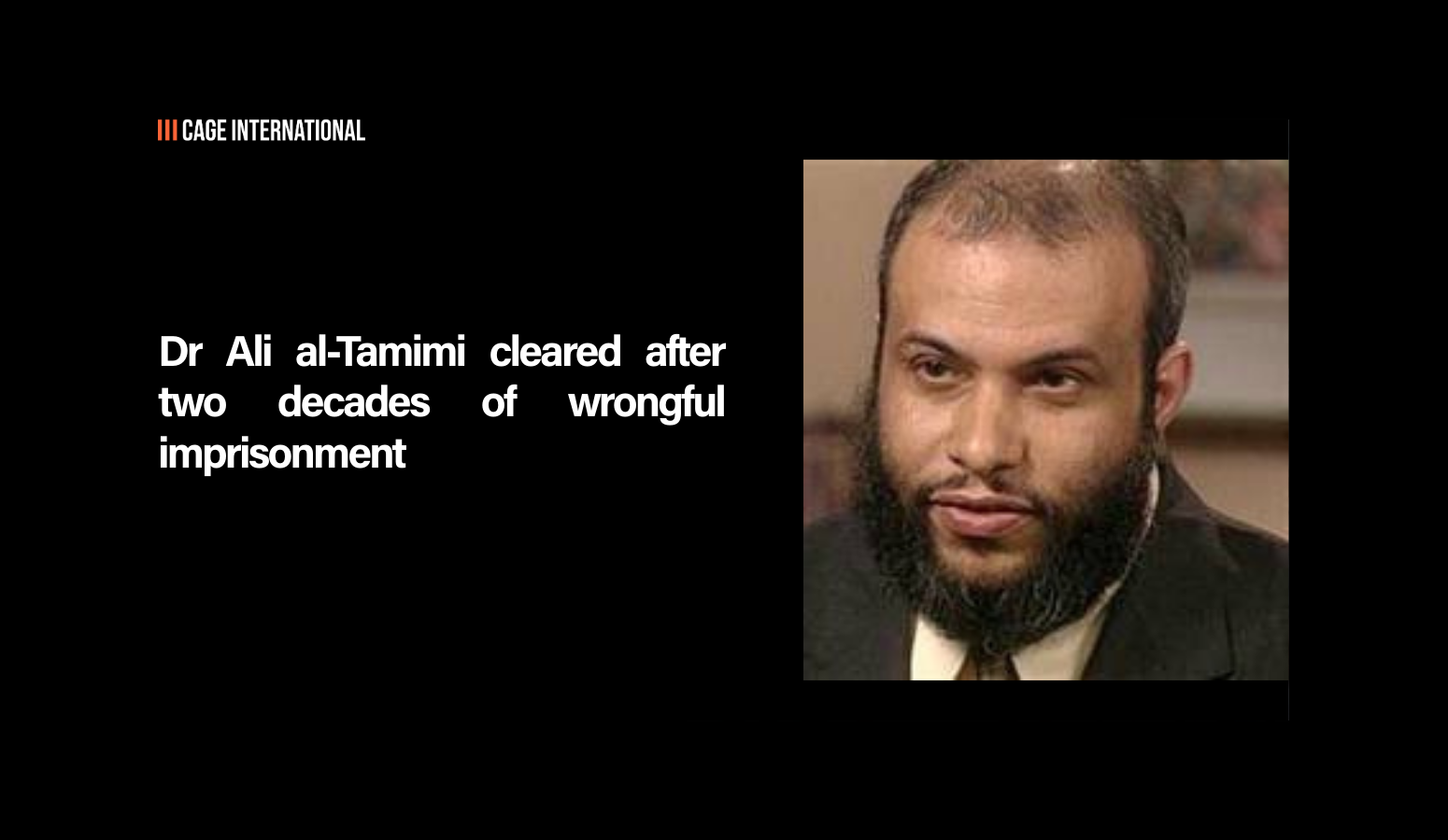
Dr Ali al-Tamimi cleared after two decades of wrongful imprisonment
January 14, 2026
OICCI session on green taxonomy highlights $565.7 billion investment needed for Pakistan’s climate goals
January 14, 2026
PCB announces schedule for three-match T20I series against Australia | Cricket News
January 14, 2026