Qaasid News
Download Our App
Latest News from Pakistan
Iran’s protests have spread across provinces, despite skepticism and concern among ethnic groups
January 13, 2026
Community pharmacy cholesterol testing pilot expands across east London – The Pharmaceutical Journal
January 13, 2026
Why the massive Iran protests haven’t toppled its clerical establishment – The Times of Israel
January 13, 2026
US urges citizens to leave Iran immediately
January 13, 2026
New publication to help small businesses manage cyber security risks from AI – Cyber.gov
January 13, 2026
Men’s Basketball Hosts Auburn on Wednesday Night
January 13, 2026
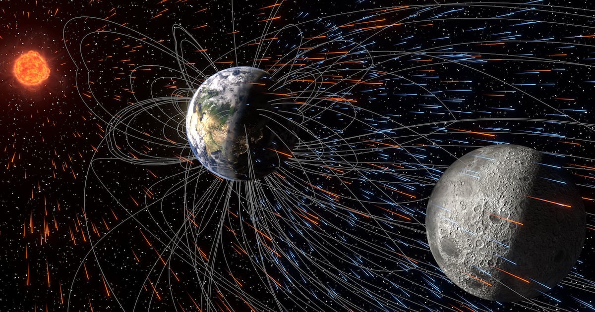
Astronomers now say the moon is eating up molecules from Earth’s atmosphere – CTV News
January 13, 2026
Jellyfish sleep and nap like us. Studying them could help human brains – NPR
January 13, 2026
Zhongping Lee awarded the Nils Gunnar Jerlov Medal
January 13, 2026
Siwarha’s Wake Gives it Away at Betelgeuse
January 13, 2026