Qaasid News
Download Our App
Latest News from Pakistan
How rising retail brands use influencers to combat digital overload
January 13, 2026
Secure Work and AI Across Any Browser
January 13, 2026
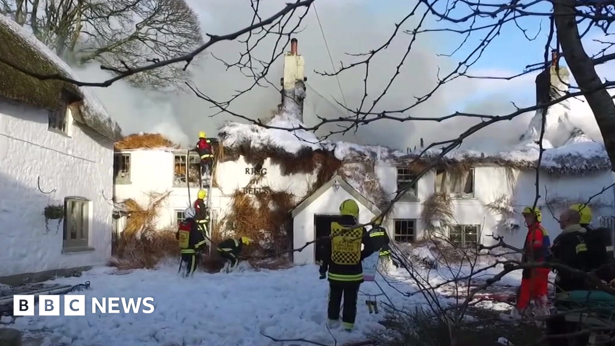
Dartmoor pub Ring of Bells’ survival after fire marked with film
January 13, 2026
Pakistan, Myanmar for resolving regional issues through dialogue – RADIO PAKISTAN
January 13, 2026
Noura Al Kaabi Meets Minister of Foreign Affairs of the Commonwealth of Dominica
January 13, 2026
4 killed in firing incident in NW Pakistan-Xinhua
January 13, 2026
4 killed in firing incident in NW Pakistan-Xinhua
January 13, 2026
NASA releases all launch dates for Artemis II. This is how soon we could be going back to the Moon
January 13, 2026
From partners to rivals: What the Saudi-UAE rupture means for Europeans – European Council on Foreign Relations
January 13, 2026
New Ambassadors present Credentials – 13 January 2026
January 13, 2026