Sample Page
Interview with Esteban Martinez: EACS Guideline updates
Written by
admin
in
6. Health
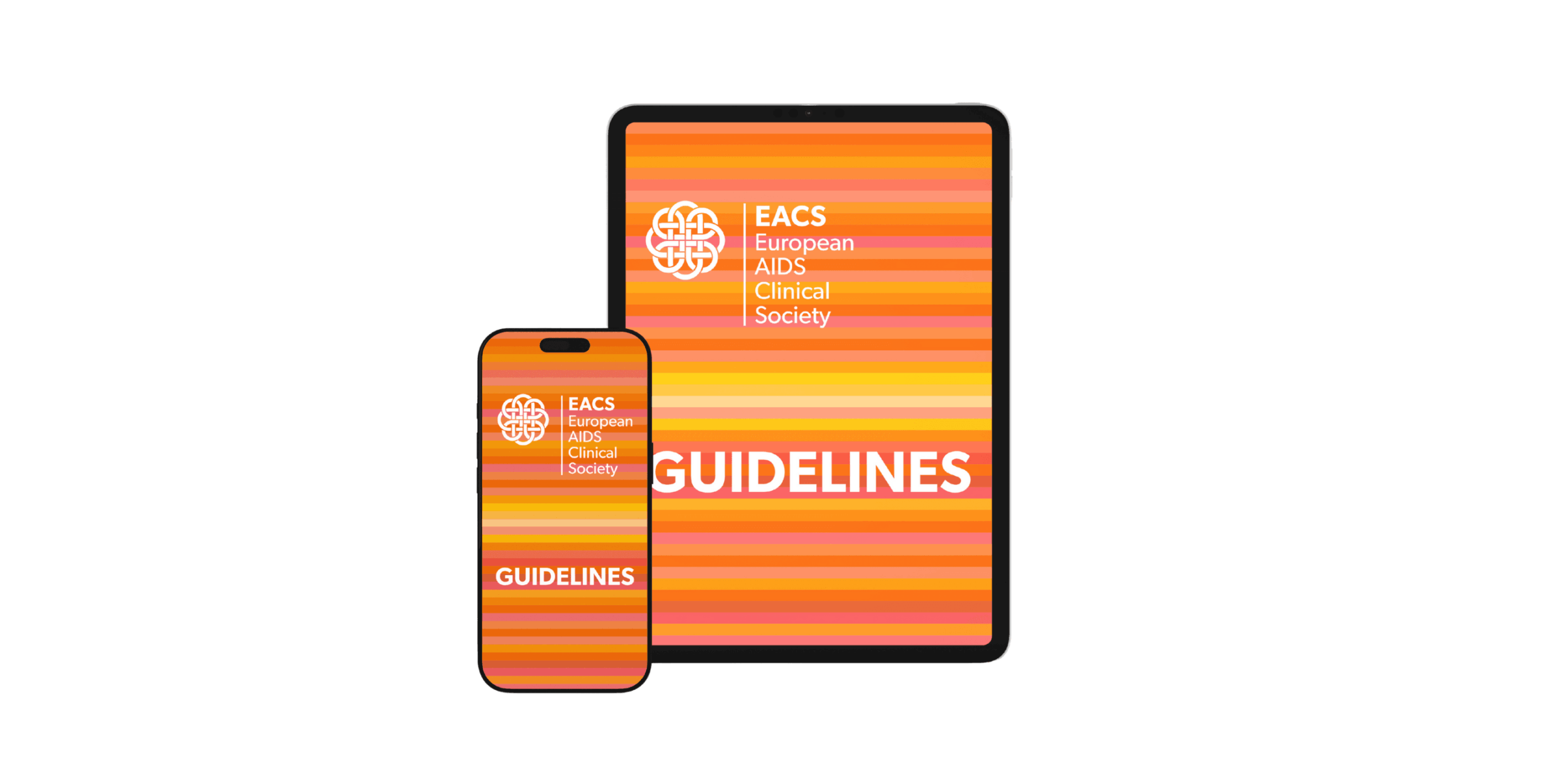
The 2025 European AIDS Clinical Society (EACS) Guidelines include major updates spanning antiretroviral therapy (ART) initiation, long-acting therapies, and comorbidity management. Which of these changes do you think will have the greatest…
Continue Reading
←
These factors appear prior to 99% of all heart attacks and strokes
How students want teachers to support them in using AI
→
More posts
Busting Prostate Cancer Myths: Facts Malaysian Men Should Know For Early Detection, Better Outcomes
December 22, 2025
MHI Group to Accelerate Development of Digital Talent– Achieving Value Creation and High Profitability Through AI and Digital Utilization —
December 22, 2025
Java News Roundup: GlassFish, TornadoVM, Spring Shell, WildFly, Hibernate, Kotlin
December 22, 2025
Engineering Microbes for Sustainable Microplastic Breakdown
December 22, 2025