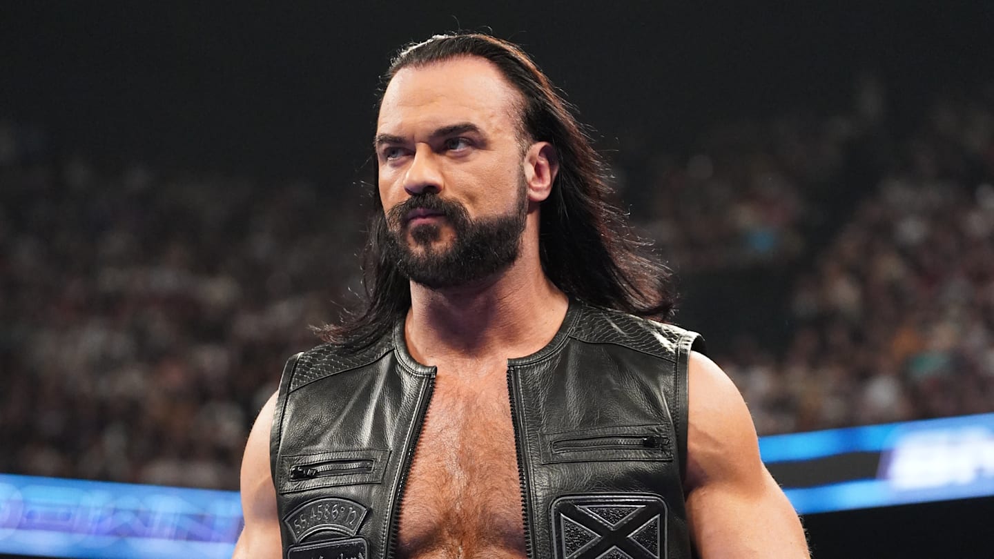
Association of alpha-1 acid glycoprotein with hepatic steatosis and liver fibrosis among women: a population-based study | BMC Gastroenterology

Our analysis of a nationally representative sample of U.S. women aged 18–49 revealed a significant positive association between AGP levels and hepatic steatosis, as measured by CAP. However, the cross-sectional design precludes determining the direction or causality of this association. For example, elevated AGP levels may coincide with or follow hepatic steatosis, possibly reflecting an inflammatory response to fat accumulation rather than playing a direct role in its development. The relationship between AGP and liver fibrosis, as assessed via LSM, is more complex. In the fully adjusted linear regression model (Model 3), no statistically significant association was observed between Ln AGP and LSM (β = −0.40, 95% CI: −0.93 to 0.12; P = 0.1307). Notably, a significant negative association was observed for the highest Ln AGP quartile (Q4) compared to Q1 in the fully adjusted model (β = −0.45, 95% CI: −0.89 to −0.01; P = 0.0442). This finding is counterintuitive, given that AGP is an acute-phase inflammatory marker that is typically associated with increased fibrosis. This unexpected finding may be due to residual confounding by unmeasured factors, such as anti-inflammatory medications commonly used in NAFLD, which might suppress AGP levels or liver stiffness. Alternatively, it could suggest compensatory mechanisms in advanced fibrosis, where chronic inflammation is associated with modified AGP expression or remodeling that lowers stiffness. These possibilities require further investigation through longitudinal studies and detailed assessments of medication use and disease stage.

Furthermore, subgroup analyses provided additional insights into the observed relationships. For the association between Ln AGP and CAP, BMI emerged as a significant moderator, with a stronger association observed among participants with a BMI of 25–29.9 kg/m² (β = 25.10, 95% CI: 11.40–38.81; P = 0.0003) and an even stronger effect in those with BMI ≥ 30 kg/m² (β = 32.89, 95% CI: 19.14–46.65; P < 0.0001), compared to a weaker association in those with BMI < 25 kg/m² (β = 6.79, 95% CI: −3.98 to 17.55; P = 0.2166). This interaction may arise from visceral adiposity, often increased in individuals with a higher BMI, promoting systemic inflammation and hepatic steatosis. As a source of pro-inflammatory cytokines, visceral fat could be linked to elevated AGP production and liver fat accumulation, explaining the stronger association in the overweight group (BMI 25–29.9 kg/m²). In contrast, for the association between Ln AGP and LSM, significant interactions were observed for smoking status, diabetes, race, and BMI, with a notably stronger positive association in participants with BMI ≥ 30 kg/m² (β = 2.40, 95% CI: 1.39–3.42; P < 0.0001) compared to no significant effect in lower BMI groups. These effects may stem from factors like smoking-induced inflammation and oxidative stress modifying fibrosis impact; diabetes-related insulin resistance and chronic inflammation altering AGP-LSM links; and race-specific genetic or socio-environmental influences on inflammatory profiles and susceptibility. Notably, the negative association in Mexican American women may reflect differences in genetics, metabolism, or healthcare access affecting AGP and fibrosis. Further research is needed to explore these mechanisms. In addition to these subgroup-specific effects, our analysis also revealed a nonlinear relationship between AGP and LSM, further complicating the interpretation of AGP’s role in liver fibrosis.

Smooth curve fitting and threshold effect analysis identified a statistically significant nonlinear (L-shaped) relationship between Ln AGP and LSM, with an inflection point at 0.05 g/L (Table 7; Fig. 3). This central finding underscores the complexity of AGP’s role in liver fibrosis. For Ln AGP < 0.05 g/L, each one-unit increase in Ln AGP was associated with a −1.16 kPa decrease in LSM (95% CI: −1.73 to −0.58; P < 0.0001). For Ln AGP > 0.05 g/L, however, a positive association emerged (β = 7.23, 95% CI: 4.76–9.71; P < 0.0001). This L-shaped pattern indicates a nonlinear threshold-dependent relationship: below the 0.05 g/L threshold, higher AGP levels are associated with reduced liver stiffness, which may reflect a quiescent inflammatory state or confounding factors. Above this threshold, however, a positive association emerges, which is consistent with AGP’s role in inflammation and fibrosis progression [9]. The inflection point at 0.05 g/L may represent a biological threshold at which AGP’s function shifts. This shift may be due to changes in glycosylation patterns or receptor interactions at higher concentrations, as observed in prior studies of liver disease [24]. Though counterintuitive for an acute-phase protein, the negative association below the threshold may align with low AGP, indicating a quiescent state, or with confounders like medication, suppressing both AGP and LSM. However, our cross-sectional design limits our ability to investigate this possibility. Furthermore, reverse causation, where advanced liver disease influences AGP expression or activity, merits consideration when interpreting this intricate association. Above the threshold, the steep positive slope aligns with AGP’s established role in inflammation and fibrosis progression [25]. These findings suggest a complex interplay between AGP and liver stiffness that potentially involves inflammatory and fibrotic pathways, which vary with AGP concentration. Further research is needed to confirm these dynamics. The nonlinear pattern may indicate differential roles of AGP in early versus advanced fibrosis stages, warranting further mechanistic and longitudinal studies to elucidate these relationships. To further explore this threshold-dependent association, we examined potential mechanisms. Medications frequently prescribed for liver disease, such as statins and non-steroidal anti-inflammatory drugs, may attenuate the AGP-LSM relationship by suppressing systemic inflammation and reducing AGP levels. This has been demonstrated in NAFLD cohorts where statin use independently lowers liver stiffness [26]. In advanced disease stages, immunosuppressive states may affect AGP production by impairing hepatic synthetic capacity and downregulating cytokine-driven acute-phase responses. This leads to decreased AGP levels, as observed in cirrhosis, where liver dysfunction correlates with reduced AGP levels. This mechanism could contribute to the negative AGP-LSM association below the threshold; however, cross-sectional data limit causal attribution. Biologically, the 0.05 g/L inflection point may signify a shift in AGP glycosylation that alters its role from protective to pro-fibrotic. Low concentrations of AGP may exert anti-inflammatory effects via immunomodulation, while higher levels may promote fibrosis through enhanced cytokine interactions [27]. This threshold-dependent change may explain the relational shift, though mechanistic details require further elucidation. Additional mechanistic pathways for the L-shaped pattern may involve oxidative stress modulation, where varying AGP levels could influence the balance of reactive oxygen species, thereby contributing to altered fibrosis dynamics [28]. Furthermore, AGP may contribute to extracellular matrix remodeling by interacting with fibrogenic factors, resulting in nonlinear stiffness changes [29]. Other pathways, including potential reverse associations in which fibrosis is associated with reduced AGP levels, underscore the need for prospective validation [30].

Our study contributes to the growing body of literature on the role of systemic inflammation in liver diseases, with a specific focus on AGP. Previous research has indicated that inflammatory markers are associated with liver pathology. For instance, studies have shown that AGP localizes in hepatocytes near fibrotic areas in chronic hepatitis, suggesting its involvement in fibrosis development [25]. However, our observation of no significant linear association between AGP and liver fibrosis, despite a nonlinear relationship, contrasts with some reports linking AGP to fibrosis progression. This discrepancy may be attributable to differences in study populations, methodologies, or disease stages. Furthermore, research indicates that AGP levels may decrease in advanced liver diseases like cirrhosis, potentially explaining the complex relationship observed in our study [30]. Alterations in AGP’s glycosylation patterns in liver diseases have also been noted, which may influence its function and association with liver outcomes [24]. These findings highlight the multifaceted role of AGP in liver pathology and underscore the need for further studies to clarify its mechanisms and clinical implications.

As far as we are aware, this study is the first to explore the association between AGP levels and hepatic fibrosis and steatosis utilizing data from the NHANES database. Previous studies on AGP in liver diseases mostly explored its potential value as a biomarker for conditions such as HCC and cirrhosis. Studies have shown that patients with liver cancer and cirrhosis exhibit diverse glycosylation alterations of AGP, including sialylation and fucosylation, compared with healthy controls [16]. Furthermore, AGP has demonstrated utility in prognostic prediction for liver disease patients. For instance, Lim DH reported that AGP, particularly its asialo form, is associated with the progression of liver diseases, suggesting its potential as a diagnostic marker for cirrhosis [12].

Currently, a substantial body of epidemiological evidence supports the pivotal role of inflammation in the progression of cirrhosis to hepatocellular carcinoma [31, 32]. In multicenter studies conducted in Italy and Finland, researchers identified a close association between histological features such as steatosis, hepatocellular ballooning, and lobular inflammation, all linked to advanced fibrosis. These findings further revealed that approximately one-third of individuals with advanced fibrosis exhibited no evidence of non-alcoholic steatohepatitis (NASH), indicating that fibrosis may develop independently of inflammation [33]. The American Association for the Study of Liver Diseases (AASLD) defines NAFLD as comprising two distinct entities: hepatic steatosis (NAFL) and NASH. NAFL is defined by hepatic steatosis of ≥ 5% without hepatocellular injury, whereas NASH is defined by the presence of steatosis, inflammation, and hepatocellular ballooning, either with or without the presence of fibrosis. These data suggest that although inflammation, specifically hepatocyte ballooning, can occur in NAFLD, fibrosis is not invariably associated with inflammation [34]. This may explain the lack of a statistically significant association between the inflammatory marker AGP and the fibrosis marker LSM in our study.

The progression from simple steatosis to NASH is closely associated with the severity of hepatic inflammation. Hepatic inflammation serves as a key factor in the progression of liver injury, subsequently exacerbating the liver’s metabolic dysfunction. Steatosis is characterized by inflammatory responses, which involve the activation of inflammatory cells, hepatocellular ballooning, and the progression of fibrosis. These events contribute to cellular damage, thereby exacerbating the severity of the condition. Histological investigations have shown that the presence of inflammation is crucial for distinguishing NASH from isolated steatosis, and it is strongly correlated with the extent of liver damage. The inflammatory environment not only accelerates steatosis but also promotes hepatocellular injury, leading to fibrosis and potentially cirrhosis [32]. Haukeland et al. investigated serum samples from 47 patients with histologically confirmed NAFLD and found that these patients typically displayed mild systemic inflammation [19]. Our study further suggests a strong positive association between AGP and CAP, indicating the potential interrelated nature of inflammation and hepatic steatosis.

NAFLD encompasses a spectrum of conditions, from benign and reversible simple steatosis to NASH, which is marked by hepatocellular injury, inflammation, and fibrosis. The transition from NAFLD to NASH is driven by metabolic dysregulation, immune activation, and chronic inflammation. Central to NASH pathogenesis is the stimulation of the innate immune system, which exacerbates hepatocellular injury and initiates pro-inflammatory pathways. This, in turn, accelerates the development of fibrosis by maintaining a persistent inflammatory response [17, 32]. This inflammation is importantly driven by major metabolic factors, including insulin resistance, excessive visceral fat accumulation, and disrupted lipid metabolism. These metabolic abnormalities lead to immune cell infiltration and the activation of inflammatory signaling pathways like the NLRP3 inflammasome, further accelerating liver injury and fibrosis. Additionally, free fatty acids (FFAs) released from visceral fat induce lipotoxicity, mitochondrial dysfunction, oxidative stress, and cytokine release, contributing to further hepatocyte ballooning and fibrosis. Recent advances in metabolomics have revealed key metabolic alterations, including the accumulation of ceramides, diacylglycerols, and FFAs, strongly linked to insulin resistance and hepatic steatosis. These metabolites not only intensify cellular stress but also activate inflammatory pathways, leading to greater liver damage and fibrosis. Notably, ceramides have been implicated in enhancing hepatic insulin resistance, promoting fibrosis, and inducing hepatocyte apoptosis [26]. Moreover, disruptions in bile acid metabolism, driven by changes in gut microbiota composition, have emerged as important regulators of liver inflammation and fibrosis. Altered bile acid profiles associate with increased inflammatory responses in the liver, underscoring the role of gut-liver interactions in NAFLD progression. Chronic liver inflammation triggers fibrotic pathways through the secretion of pro-inflammatory cytokines, including TNF-α and IL-1β, which activate hepatic stellate cells. This stimulation triggers a series of events that lead to collagen buildup and fibrosis progression [35].

This study has several critical limitations that must be considered. Chief among them is the cross-sectional design, which inherently prevents the establishment of temporal relationships or causal inference. Consequently, our findings should be interpreted strictly as associations, and we cannot determine whether AGP influences liver outcomes such as hepatic steatosis or fibrosis, or if these liver conditions lead to elevated AGP levels. The absence of longitudinal data precludes us from assessing temporality, and the possibility of reverse causality—where hepatic conditions, such as steatosis, may elevate AGP levels rather than AGP contributing to liver pathology—cannot be excluded. Although we adjusted for several potential confounders, residual confounding remains a concern, particularly given the lack of data on medication use and other unmeasured factors that could influence both AGP and liver health. Specifically, medications such as steroids, non-steroidal anti-inflammatory drugs (NSAIDs), and statins—known to modulate AGP levels due to their effects on inflammation or lipid metabolism—were not included in our analysis, despite being collected in the NHANES database, likely introducing residual confounding. Moreover, genetic factors, such as the well-established variants PNPLA3 and TM6SF2, which are associated with increased hepatic fat accumulation and fibrosis risk, may also correlate with systemic inflammation and AGP levels; these were not accounted for in our study. Furthermore, as an acute-phase protein, AGP is subject to diurnal fluctuations and acute elevations triggered by recent illness, infection, or psychological stress—variables that were neither adjusted for nor excluded in our analysis—potentially compromising the consistency and reliability of AGP measurements. Second, the final study population consisted solely of women aged 18–49 years, an outcome of the exclusion criteria and data availability rather than an intentional restriction. While this focus enabled a detailed analysis within this group, it limits the generalizability of our findings to men, older women, or younger individuals. Third, despite adjustments for several potential confounders, the possibility of residual confounding persists, particularly due to the lack of detailed information on medication use in the NHANES database. Anti-inflammatory medications, commonly prescribed to individuals with NAFLD, could influence AGP levels and were not accounted for in our analysis. Fourth, hepatic steatosis and liver fibrosis were evaluated using transient elastography, a validated non-invasive technique. However, while transient elastography demonstrates high accuracy, it is not a substitute for liver biopsy, and its performance has not been specifically validated in our study population. Additionally, there is a potential for false positives or false negatives, particularly in individuals with high BMI or other factors that may affect measurement accuracy. Therefore, our findings may differ from those obtained through histological assessment. Fifth, the absence of ethnicity-specific stratified analyses represents a significant limitation, given the documented variability in AGP concentrations across ethnic groups, which may reflect differences in genetic backgrounds or inflammatory profiles. This omission may reduce the applicability of our findings to diverse populations with distinct AGP baselines. It is important to acknowledge that in the context of multiple comparisons, such as those performed in subgroup analyses, findings with p-values approaching but not meeting the conventional threshold for statistical significance should be interpreted with particular caution due to the potential for limited statistical power in smaller subgroups and an increased possibility of false positive results. Finally, the exclusion of a large portion of the initial sample due to missing data may introduce selection bias, potentially affecting the representativeness of our results. Nevertheless, our study presents several strengths. By utilizing a nationally representative sample, our results can be generalized to a broad adult population in the United States, representing various ethnicities and gender groups. Additionally, the extensive sample size enabled subgroup analyses, enhancing the robustness of our results.

Continue Reading