Written by
in
Published on December 7, 2025
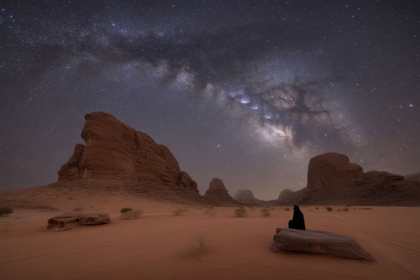
Astronomy is experiencing a remarkable renaissance in Saudi Arabia’s vast…