New evidence counters the conventional wisdom that limb amputation leads to reorganization of the map the brain uses to track the body’s location. When researchers compared three patients before and after hand amputation, they found the somatosensory cortex remained largely the same.
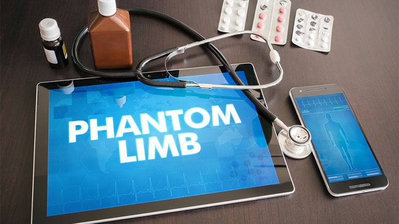
These findings point to new approaches to the management of phantom limb pain, a debilitating condition with a lifetime prevalence of 76%-87% after amputation.
Rather than relying on common rehabilitation strategies like mirror box therapy, virtual reality, or graded motor imagery, reconnecting severed nerves to muscle or other tissues may reduce the risk for phantom pain. These findings could also inform the redesign of brain-computer interfaces.
“The take-home is that the brain’s body map remains highly preserved after amputation,” lead study author Hunter Schone, PhD, postdoctoral associate in the Rehab Neural Engineering Labs at the University of Pittsburgh, told Medscape Medical News. “Our results show…the sensory map is maintained, with no evidence for reorganization.”
The study was published online on August 21 in Nature Neuroscience.
Long-Standing Theory Upended
The current research also counters a long-standing belief that cortical maps are competitive. Traditional thinking is that when one input disappears, neighboring inputs expand to take over the area changed through amputation. He and colleagues refuted this by tracking input from the lips, which did not take over the parts of the brain map that normally track the hand and fingers.
Unlike previous cross-sectional studies, the researchers in this study scanned the same patients both before and after planned amputations. This design allowed testing this competitive belief directly for the first time, added Schone, who is also affiliated with the National Institutes of Health’s Laboratory of Brain and Cognition, National Institute of Mental Health in Bethesda, Maryland, and the Institute of Cognitive Neuroscience, University College London, London, England.
“Across multiple analyses, we saw no evidence of takeover,” Schone said. “These findings call for a critical rethinking of phantom limb pain treatments, shifting focus downstream, toward peripheral nerves and the spinal cord.” The investigators assessed the three adults before and 6 months after hand amputation. The procedure was indicated because of a rapidly developing arteriovenous malformation in the upper arm of one patient, to remove a slow-growing sarcoma tumor in a second, and because of multiple bone fractures in an arm resulting from Servelle-Martorell syndrome in the third participant.
A control group of 16 able-bodied individuals was included in the study. An additional cross-sectional analysis involved amputees and controls from three previous studies.
Participants completed functional neuroimaging tests, including finger motor control, kinesthetic vividness, and a functional index, as well as pain rating scales. Specific tasks included visually cued finger tapping, lip pursing, and toe flexing during scanning. Researchers created contrast maps for attempted vs phantom hand movements, for hand and lip movements, and for finger selectivity.
The results showed “strikingly consistent hand and lip cortical maps” before and following surgery, the researchers noted.
The hand and individual finger activity in the somatosensory cortex remained stable. Phantom activity was similar in amplitude and spatial activity spread before surgery.
“We were struck by the remarkable stability of the hand map, even after years without the hand’s rich sensory input to the brain,” Schone said. The hand map did not fade over time. Instead, it retained such precision that a machine learning decoder trained on pre-amputation finger movements reliably decoded phantom finger movements years later.
“This degree of selectivity after such a dramatic loss of input was unexpected,” Schone added.
The investigators also compared their case studies with a group of 26 chronic upper-limb amputees an average of 23.5 years after surgery. The authors noted that the similar findings suggest hand and lip representations remain stable over the long term, even after amputation.
Future Research Goals
Schone and colleagues plan to investigate why the brain maintains these maps in the absence of the sensory input. This persistence suggests that the somatosensory cortex is more than a passive relay of signals from the periphery, Schone said.
The researchers also plan to use these findings to develop next-generation brain-computer interfaces. The goal is to take advantage of these preserved maps to restore movement and sensation for people with amputations or spinal cord injuries.
“Because the adult brain maintains these sensory-deprived representations, they can serve as a stable foundation for clinical translation of these technologies,” Schone added.
The study was independently supported. Schone reported having no relevant financial relationships.