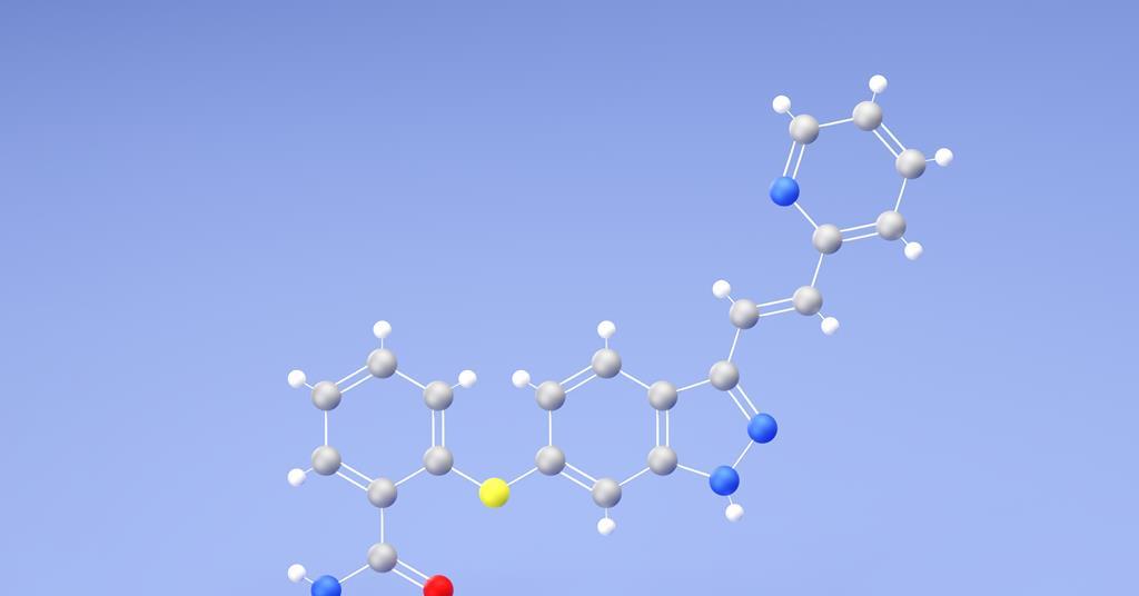
Microbial factors associated with HPV infection and viral clearance: i

Introduction

Cervical cancer (CC) is one of the most common malignant tumors affecting women worldwide, with human papillomavirus (HPV) infection recognized as its necessary but insufficient cause. Persistent infection with high-risk HPV (HR-HPV), particularly types such as HPV16 and HPV18, plays a pivotal role in the development of cervical intraepithelial neoplasia (CIN) and CC.1 According to the World Health Organization, over 600,000 new CC cases and more than 340,000 deaths occur annually, with the highest burden concentrated in low- and middle-income countries (LMICs), particularly in sub-Saharan Africa, Southeast Asia, and Latin America.2 These global disparities are influenced not only by differences in screening and vaccination coverage but also by variations in vaginal microbiota composition and host immune responses across populations.3 However, only a small proportion of HPV infections progress to malignancy, suggesting that host immunity, viral genotype, and the cervicovaginal microenvironment critically influence the natural history of HPV infection.4 As HPV vaccine uptake and screening programs advance in high-income countries, understanding the microbiological and immunological drivers of HPV progression in diverse global populations has become increasingly urgent for eliminating CC as a public health problem.

The vaginal microecosystem is a complex, dynamic environment composed of microbial communities, immune factors, hormones, and metabolites. In healthy reproductive-age women, this microenvironment is usually dominated by Lactobacillus species such as L. crispatus, L. gasseri, L. iners, and L. jensenii, which help maintain acidic pH, produce hydrogen peroxide (H2O2), and compete against pathogenic microbes.5 Disruption of this balance, known as vaginal dysbiosis, is characterized by a depletion of Lactobacillus and a concurrent increase in anaerobic bacteria such as Gardnerella, Atopobium, and Prevotella. Over the past decade, increasing global evidence has indicated that disturbances in vaginal microecology and associated immune dysregulation play critical roles in HPV susceptibility and persistence,6,7 and progression to cervical disease.8 Worse still, this abnormal condition is associated with increased susceptibility to sexually transmitted infections, including HPV.9,10 For instance, bacterial vaginosis (BV)—a common dysbiotic state—has been consistently associated with reduced HPV clearance rates and elevated CIN risk.11 It is inferred that BV-associated bacteria and their metabolites, including sialidase and polyamines, may compromise mucosal barrier function and alter local immune responses, facilitating viral persistence.12 Moreover, an imbalance in microbiota can lead to altered production of cytokines such as IL-6, IL-8, and TNF-α, which are linked to chronic inflammation and impaired viral clearance.13 Studies from Africa, Asia, and Europe confirm that women with non-Lactobacillus-dominated microbiota are at higher risk of HPV persistence and progression to high-grade cervical lesions.4,14,15 These findings underscore the importance of understanding microbe-host-HPV interactions across different populations.

Hydrogen peroxide-producing Lactobacillus species, in particular, appear protective against HPV. These microbes can modulate immune responses, reduce local inflammation, and inactivate viruses via oxidative stress.16 Conversely, reduced H2O2 levels have been observed in HPV-infected individuals, suggesting a compromised microbial defense.17 Furthermore, recent evidence has shown that HPV infection itself may downregulate mucosal innate peptides used by Lactobacilli for growth, thereby reinforcing dysbiosis and perpetuating viral persistence—a novel mechanism of immune evasion.6 Furthermore, cytokines such as IL-6 and IL-12 play roles in antiviral defense and T-cell recruitment. Studies have shown that decreased IL-12 and elevated IL-6 levels in the vaginal milieu may impair effective immune responses, leading to persistent HPV infections.18 Additionally, factors such as vaginal pH, leukocyte esterase (LE) activity, clue cells, and overall cleanliness score serve as indirect markers of local immunity and microbial composition, and may provide clinical clues to HPV infection status.19 Thus, it is reasonable to pay more attention to the factors potentially modulating the abundance of Lactobacilli and immune state in vaginal microecosystem during the management of HPV infections.

Despite accumulating evidence regarding the HPV infection and dysbiosis of vaginal microbiome, most prior studies are either cross-sectional or based on small sample sizes. Moreover, few have integrated microbial, immunological, and HPV genotyping data longitudinally. There remains a need for real-world, retrospective analyses that explore the interaction between vaginal microecological characteristics and HPV persistence and clearance over time. The clarification of these relationships is essential for developing predictive models, identifying high-risk patients, and optimizing early interventions.

Thus, in the current study, we performed a two-year retrospective study involving 312 women, which aimed to investigate the correlation between vaginal microecological status and HPV infection comprehensively, with a focus on identifying microbial and biochemical predictors of viral persistence or clearance. By integrating pH, cleanliness grade, enzyme activity (H2O2, LE, sialidase), and HPV genotyping data, the current study provides new insights into how host-microbe-viral interactions influence the natural course of HPV infection. Our findings may provide additional information regarding the risk stratification of CC, and guide targeted strategies to enhance HPV clearance in additional to the previous publications.

Methods

Participants

The current study is a retrospectively observational study, which conducted at the Fifth Hospital of Xiamen based on records collected between January 2023 and December 2024. Eligible participants were women aged 20 to 65 years who underwent comprehensive gynecological screening, including HPV genotyping (performed using the Hybribio 21 HPV Genotyping Assay; DNA extracted with Qiagen QIAcube and PCR run on ABI 7500 Real-Time PCR System), vaginal microecology assessment, cytological evaluation, and biochemical analysis of vaginal secretions. Inclusion criteria were: (1) availability of baseline and follow-up HPV testing, (2) standardized and complete vaginal microecological testing, and (3) complete demographic and clinical records. Exclusion criteria included prior treatment for cervical neoplasia, history of cervical surgery, immunosuppression (eg, HIV infection, corticosteroid use), pregnancy during the study period, antibiotic or probiotic use within four weeks before sampling, and incomplete data. Ethical approval was granted by the Fifth Hospital of Xiamen, and the study adhered to the Declaration of Helsinki guidelines. All the patients have signed an informed consent regarding the use of clinicopathological information prior to study commencement.

Clinical and Laboratory Data Collection

Demographic information (age, parity, smoking status, contraception method), clinical examination findings, and results of HPV genotyping, vaginal microecology, cytology, and inflammatory marker detection were retrieved from the hospital information system by trained researchers. Data were cross-verified independently to ensure accuracy.

HPV Detection and Genotyping

Cervical samples were collected using sterile cytobrushes and stored in liquid-based cytology medium. HPV DNA extraction was performed using a commercial DNA extraction kit following standardized protocols. HPV genotyping was conducted using a multiplex PCR assay (ABI 7500 Real-Time PCR System, USA), which is capable of identifying 14 high-risk types (including HPV 16, 18, 31, 33, 45, 52, and 58) and 5 low-risk types (HPV 6, 11, 42, 43, and 44). Where available, viral load was inferred from Ct values, with Ct <30 indicating high viral burden, 30–35 moderate, and >35 low viral burden. Infection outcomes were categorized as clearance, persistence, or progression, depending on genotype detection across serial timepoints.

Vaginal Microecology Evaluation

Vaginal secretions were sampled before pelvic examination using sterile cotton-tipped applicators. Samples were immediately evaluated for multiple parameters. pH was measured using narrow-range indicator strips (precision 0.1 pH units), and values ≥4.5 were considered elevated, indicating potential dysbiosis. Cleanliness was graded via a phase-contrast microscopy (Leica DM750, Germany). (400× magnification) into four grades: Grade I (predominant Lactobacillus, rare leukocytes), Grade II (moderate Lactobacillus with occasional mixed flora), Grade III (mixed flora, moderate leukocytes), and Grade IV (predominance of pathogenic flora, heavy leukocytes).

Gram-stained smears were subjected to Nugent scoring: (1) Lactobacillus morphotype (large Gram-positive rods), (2) Gardnerella morphotype (small Gram-variable rods), and (3) Mobiluncus morphotype (curved Gram-negative rods). Nugent scores were stratified into normal (0–3), intermediate (4–6), and bacterial vaginosis (7–10). Furthermore, samples were subclassified into “low-intermediate” (4–5) and “high-intermediate” (6) groups to capture finer gradations.

Clue cells, Candida spp., and Trichomonas vaginalis were identified by a phase-contrast microscopy (Leica DM750, Germany). The proportion of clue cells was recorded (% of epithelial cells affected), with >20% considered significant. Budding yeast and pseudohyphae indicated fungal infection; motile flagellated organisms were diagnostic of trichomoniasis.

Hydrogen peroxide (H2O2) production was assessed semi-quantitatively by colorimetric testing and graded as strong, weak, or negative using a microplate reader (Thermo Fisher Scientific, USA). Weak or negative production suggested impaired Lactobacillus function. Lactobacillus subtype identification (where available) was performed by culture and biochemical profiling, differentiating between L. crispatus, L. iners, L. gasseri, and L. jensenii species. L. crispatus predominance was considered indicative of optimal microecology, while L. iners predominance was classified as intermediate.

Biofilm formation on epithelial cells was evaluated qualitatively via Gram staining and categorized as absent, sparse, moderate, or dense biofilm layer, particularly noting Gardnerella or Atopobium dominance.

Leukocyte esterase (LE) activity was measured by urinary dipstick on vaginal secretions: LE positivity (≥1+) indicated local inflammatory activation. Vaginal epithelial integrity was evaluated by the presence of parabasal cells, indicating atrophic change.

Sialidase enzyme activity was detected using a colorimetric rapid detection kit under a microplate reader (Thermo Fisher Scientific, USA), indicating enzymatic disruption of epithelial barriers associated with BV pathogens.

Community state types (CSTs) were determined based on dominant flora patterns: CST I (L. crispatus-dominant), CST II (L. gasseri-dominant), CST III (L. iners-dominant), CST IV (diverse anaerobes), and CST V (L. jensenii-dominant).

Definitions

Normal vaginal microecology was defined as pH <4.5, Nugent score 0–3, H2O2 strong positive, dominant Lactobacillus spp. (preferably L. crispatus or L. jensenii), absence of biofilm and clue cells, and negative LE and sialidase tests. Dysbiosis was defined as pH ≥4.5, Nugent score ≥4, decreased or absent H2O2, dominance of anaerobes (eg, Gardnerella, Atopobium, Prevotella), positive LE or sialidase, and evidence of biofilm formation. HPV persistence was defined as detection of the same HPV genotype across two consecutive tests ≥12 months apart.

Cytological Examination

Cervical cytology specimens were collected via the ThinPrep® method, fixed in PreservCyt® solution, and processed within 7 days. Slides were reviewed by two independent cytopathologists according to the 2014 Bethesda System, classifying samples as negative for intraepithelial lesion or malignancy (NILM), atypical squamous cells (ASC-US, ASC-H), low-grade squamous intraepithelial lesion (LSIL), high-grade squamous intraepithelial lesion (HSIL), or atypical glandular cells (AGC). All disagreements were resolved by consensus conference.

Biochemical and Inflammatory Marker Detection

For a subset of patients, cervicovaginal secretions were analyzed for soluble immune and inflammatory markers. Concentrations of interleukin-6 (IL-6) (H007-1-1, Nanjing Jiancheng Bioengineering Institute, China), interleukin-12 (IL-12) (H010-1-2, Nanjing Jiancheng Bioengineering Institute, China), tumor necrosis factor-alpha (TNF-α) (H052-1-2, Nanjing Jiancheng Bioengineering Institute, China), and secretory immunoglobulin A (sIgA) (H108-2-1, Nanjing Jiancheng Bioengineering Institute, China) were measured using high-sensitivity enzyme-linked immunosorbent assay (ELISA) kits following standard protocols. Systemic inflammation was assessed by measuring C-reactive protein (CRP) levels in serum samples where available (E023-1-1, Nanjing Jiancheng Bioengineering Institute, China). Additionally, oxidative stress markers, including malondialdehyde (MDA) levels, were measured in vaginal fluids using thiobarbituric acid reactive substances (TBARS) assays (A003-1-2, Nanjing Jiancheng Bioengineering Institute, China).

Statistical Analysis

All statistical analyses were conducted using SPSS version 26.0 (IBM Corp., Armonk, NY, USA) and R software version 4.2.1. Continuous variables were assessed for normality using the Kolmogorov–Smirnov test. Normally distributed variables were presented as mean ± standard deviation (SD) and compared using independent-sample t-tests; non-normally distributed variables were expressed as median (interquartile range, IQR) and compared using the Mann–Whitney U-test. Categorical variables were compared using the Chi-square test or Fisher’s exact test, as appropriate. Multivariate logistic regression was performed to identify independent predictors of HPV persistence and clearance, adjusting for potential confounders. Odds ratios (ORs) and 95% confidence intervals (CIs) were reported. ROC curve analysis was performed to assess the predictive power of key parameters, and the area under the curve (AUC) was calculated. Kaplan–Meier survival curves were generated to compare HPV clearance rates between different vaginal microecology groups, and statistical significance was assessed using the Log rank test. A two-sided P value <0.05 was considered statistically significant.

Results

Baseline Characteristics

A total of 312 women were included in the final analysis cohort. As shown in Table 1, the mean age of the participants was 38.7 ± 9.6 years. Most women were multiparous (68.6%), and the majority used condoms as their primary contraceptive method (42.9%), while 10.9% reported smoking habits. Baseline HPV positivity was observed in 210 cases (67.3%), and elevated vaginal pH (≥4.5) was detected in 59.0% of the population (Table 1). The bacterial vaginosis, defined by a Nugent score ≥7, was present in 28.2% of all the case (Table 1). Half of the women exhibited positive hydrogen peroxide production (50.0%), while leukocyte esterase and sialidase activities were positive in 35.9% and 23.7%, respectively (Table 1). L. crispatus was the predominant strain in 39.1% of cases, and CST I (L. crispatus-dominant) accounted for 35.3% of the vaginal community structures. Biofilm formation was noted in 33.3% of cases. Other parameters including cytokine levels, oxidative stress markers, and inflammatory indicators are detailed shown in Table 1.

Table 1 Baseline Characteristics of the Study Population

Analysis of Vaginal Microecological Status of the Study Population

Vaginal microecological parameters significantly differed between HPV-negative and HPV-positive groups. Women with HPV infection exhibited a significantly higher rate of elevated vaginal pH (76.2% vs 23.5%, P<0.001), increased prevalence of bacterial vaginosis (37.1% vs 9.8%, P<0.001), and reduced hydrogen peroxide production (41.0% vs 68.6%, P<0.001) compared to HPV-negative women (Table 2). Similarly, leukocyte esterase positivity and sialidase activity were significantly more common among HPV-positive participants (Table 2). Furthermore, CST IV (diverse anaerobic flora) predominated in HPV-positive individuals (34.3% vs 13.7%, P<0.001), and biofilm formation was substantially more frequent (41.9% vs 15.7%, P<0.001) (Table 2).

Table 2 Vaginal Microecological Characteristics Stratified by Human Papillomavirus (HPV) Infection Status

Analysis of Immune and Inflammatory Markers of the Study Population

Among the 210 HPV-positive women, 132 (62.9%) cleared the infection during the follow-up, 66 (31.4%) exhibited persistent infection, and 12 (5.7%) progressed to higher-grade cervical lesions (Table 3). Cytokine profiling revealed that women with persistent or progressive HPV infections had significantly higher IL-6 levels compared to those who cleared the infection (Persistence: 10.5 pg/mL vs Clearance: 6.8 pg/mL; Progression: 14.2 pg/mL, P<0.001) (Table 3). Conversely, IL-12 levels were significantly lower in persistence and progression groups compared to clearance (Persistence: 10.2 pg/mL vs Clearance: 14.3 pg/mL, P<0.001) (Table 3). TNF-α levels and oxidative stress marker MDA were also elevated in persistent and progression groups (Table 3). Additionally, CRP levels were higher and sIgA levels lower among women who failed to clear HPV (Table 3).

Table 3 Comparison of Immune and Inflammatory Markers Between Human Papillomavirus (HPV) Clearance, Persistence, and Progression Groups

Multivariate Analysis for Predictors of HPV Persistence

Multivariate logistic regression identified several independent risk factors associated with HPV persistence (Table 4). Elevated vaginal pH (OR=2.61, 95% CI: 1.52–4.50, P<0.001), bacterial vaginosis (Nugent score ≥7) (OR=3.23, 95% CI: 1.75–5.96, P<0.001), negative hydrogen peroxide production (OR=2.87, 95% CI: 1.54–5.35, P=0.001), and positive sialidase activity (OR=3.18, 95% CI: 1.67–6.04, P<0.001) were all significantly associated with persistence. Furthermore, CST IV (compared to CST I) (OR=2.91, 95% CI: 1.47–5.75, P=0.002), biofilm presence (OR=2.45, 95% CI: 1.32–4.57, P=0.005), and elevated IL-6 levels (>9 pg/mL) (OR=3.05, 95% CI: 1.62–5.75, P=0.001) also independently predicted persistent infection (Table 4).

Table 4 Multivariate Logistic Regression Analysis for Predictors of Human Papillomavirus (HPV) Persistence

Predictive Performance of Microecological and Inflammatory Markers

ROC analysis based on multivariate analysis demonstrated that IL-6 levels provided the highest predictive value for HPV clearance among single markers (AUC = 0.789, 95% CI: 0.721–0.856), followed closely by Nugent score (AUC = 0.778) and vaginal pH (AUC = 0.752) (Table 5). Hydrogen peroxide production and sialidase activity also showed moderate predictive accuracy. Importantly, a combined predictive model incorporating microecological and immune markers yielded the highest AUC value (0.842, 95% CI: 0.780–0.904), significantly improving sensitivity and specificity (Table 5).

Table 5 Receiver Operator Curve Analysis for Predictive Markers of Human Papillomavirus (HPV) Clearance

Further, HPV clearance rates over time differed significantly between women with normal vaginal microecology and those with dysbiosis. At 24 months, 90.1% of women with normal microecology cleared HPV compared to only 66.2% of those with dysbiosis (P<0.001) (Table 6).

Table 6 Human Papillomavirus (HPV) Clearance Rates at Different Follow-up Points Based on Vaginal Microecology Status

Discussion

The current study retrospectively explored the association between vaginal microecological status and HPV infection outcomes among 312 women over a two-year period. The findings revealed that disturbances in the vaginal microbiota, notably elevated pH, BV, decreased H2O2 production, presence of biofilms, and shifts towards diverse anaerobic CST IV, were strongly associated with HPV persistence and progression. Furthermore, inflammatory markers, particularly elevated IL-6 levels and oxidative stress markers such as MDA, emerged as significant predictors of persistent HPV infection.

Dysbiosis, characterized by increased vaginal pH and BV, predisposed to HPV persistence aligns with earlier reports emphasizing the critical role of a Lactobacillus-dominant environment in maintaining mucosal immunity.4,8,9 Previous longitudinal studies have similarly demonstrated that women with a Lactobacillus-depleted microbiota, especially those dominated by Gardnerella, Atopobium, or Prevotella, are at higher risk for HPV acquisition and lower clearance rates.20,21 In particular, CST IV, characterized by anaerobic dominance, was significantly associated with persistent infection in our cohort, corroborating previous observations.22 H2O2-producing Lactobacillus species, notably L. crispatus, have been shown to provide a protective effect against HPV persistence through direct antiviral activities and maintenance of low vaginal pH.23 Our finding that reduced H2O2 production independently predicted persistence supports this mechanistic model. Furthermore, sialidase activity, a marker of BV-associated bacteria, emerged as a strong predictor, consistent with its role in disrupting epithelial barriers and modulating local immune responses unfavorably.24 The immune microenvironment appeared equally crucial. Elevated IL-6 and reduced IL-12 levels among women with persistent or progressive HPV infection reflect a shift towards a pro-inflammatory, but ineffective antiviral state.12,25,26 IL-6 is known to promote chronic inflammation and immune evasion by HPV, while IL-12 is essential for promoting cytotoxic T-cell responses necessary for viral clearance.27 Similar cytokine profiles have been reported in earlier cross-sectional studies and CC precursor lesion studies.28–30 These findings directly address our primary research question: whether specific vaginal microecological features and immune markers are predictive of HPV persistence or clearance. The results validate our hypothesis that a dysregulated vaginal environment characterized by Lactobacillus depletion, elevated pH, enzymatic imbalances, and altered cytokine profiles are critical contributors to persistent HPV infection. By systematically integrating microbiota composition, biochemical markers, and immune parameters, the study moves beyond simple associations to propose a more comprehensive model of HPV pathogenesis.

The ROC analysis demonstrated that while individual microecological markers such as pH, Nugent score, and H2O2 status had moderate predictive value, the combination of microecological and immune parameters significantly improved the prediction of HPV clearance. This finding highlights the necessity of an integrated approach in risk stratification for women with HPV infection, an aspect underappreciated in many previous reports.31 The presence of biofilm, frequently associated with Gardnerella vaginalis and Atopobium vaginae, was also independently associated with persistence. Biofilms can shield pathogenic bacteria from host immunity and antibiotics, contributing to chronic infections and sustained mucosal inflammation.32 Recent advanced microscopy studies have confirmed the existence of polymicrobial biofilms in women with BV and HPV co-infection, supporting our findings.33 Our findings are consistent with previously international studies. A large European cohort study by Mitra et al found that a Lactobacillus-depleted, diverse vaginal microbiota significantly correlated with CIN2+ development across multiple populations.8 Similarly, Brotman et al in the United States demonstrated that changes in vaginal microbiota precede HPV detection, underscoring causality.21 In Sub-Saharan Africa, Happel and et al observed that women with high-diversity vaginal communities were more likely to harbor persistent high-risk HPV genotypes.34 These global findings corroborate our results and underscore the widespread relevance of vaginal microbiome profiling in HPV management. Hence, the significance of our study extends beyond national boundaries and offers implications for diverse populations.

Clinically, our results emphasize the importance of considering vaginal microecological assessments as part of HPV management strategies. Women with evidence of dysbiosis or elevated inflammatory markers may benefit from closer surveillance or interventions aimed at restoring healthy vaginal microbiota. Probiotic therapies, particularly those using L. crispatus strains, have shown promise in small trials for BV treatment and may warrant exploration in the context of HPV infection.35 However, several limitations must be acknowledged. First, the current study was retrospective and single-centered, which may introduce selection bias. Second, although we assessed a wide range of microecological and immune parameters, other factors such as sexual behavior, co-infections with other sexually transmitted infections, and host genetic susceptibility were not analyzed. Third, cytokine measurements were performed on cervicovaginal lavage rather than tissue samples, which may not fully reflect tissue-level immune dynamics. Future studies should focus on longitudinal interventional designs to determine whether correcting vaginal dysbiosis or modulating immune responses can actively promote HPV clearance. Advanced sequencing methods such as shotgun metagenomics may also provide deeper insights into the microbial community functional profiles associated with HPV outcomes.36 Furthermore, integration of systemic markers such as plasma cytokine levels and host immunogenetic profiling could refine predictive models.

Conclusions

In conclusion, our retrospective study uniquely integrated microbial enzymatic markers, vaginal ecological indicators, and HPV genotyping to explore the interplay between vaginal microenvironmental factors and HPV infection risk. The novelty of this work lies in its real-world clinical dataset, comprehensive evaluation of non-invasive biochemical parameters, and combined predictive value of vaginal microecology and immune markers, which were rarely analyzed together in prior studies. Our findings highlights that specific vaginal microecological disturbances are associated with HPV positivity, underscoring the diagnostic and preventive value of assessing host–microbe–virus interactions in clinical practice. However, our study is limited by its retrospective design, single-center sample, and absence of metagenomic or immunological profiling, which restricts causal inference and generalizability. Unmeasured behavioral or demographic factors may also confound the observed associations. Even though for these limitations, interventional studies targeting the vaginal microbiome may offer new strategies for enhancing HPV clearance and preventing cervical disease progression. Future research should incorporate prospective, multi-center cohorts and apply integrated multi-omics approaches to deepen understanding of host-microbiome-HPV interactions.

Data Sharing Statement

The data will be provided by the corresponding author on reasonable request.

Statement of Ethics

All the investigation of the current study was performed under the approval of the ethic committee of the Fifth Hospital of Xiamen as well as the Declaration of Helsinki.

Author Contributions

All authors made a significant contribution to the work reported, whether that is in the conception, study design, execution, acquisition of data, analysis and interpretation, or in all these areas; took part in drafting, revising or critically reviewing the article; gave final approval of the version to be published; have agreed on the journal to which the article has been submitted; and agree to be accountable for all aspects of the work.

Funding

This study is supported by Guiding Project of Medical and Health Care in Xiamen City (No. 3502Z20209233).

Disclosure

The authors disclose no conflicts of interest in this work.

References

1. Walboomers JM, Jacobs MV, Manos MM, et al. Human papillomavirus is a necessary cause of invasive cervical cancer worldwide. J Pathol. 1999;189(1):12–19. doi:10.1002/(sici)1096-9896(199909)189:1

2. Organization WH. Cervical cancer. 2022.;

3. Serrano B, Brotons M, Bosch FX, Bruni L. Epidemiology and burden of HPV-related disease. Best Pract Res Clin Obstet Gynaecol. 2018;47:14–26. doi:10.1016/j.bpobgyn.2017.08.006

4. Usyk M, Zolnik CP, Castle PE, et al. Cervicovaginal microbiome and natural history of HPV in a longitudinal study. PLoS Pathog. 2020;16(3):e1008376. doi:10.1371/journal.ppat.1008376

5. Chen X, Lu Y, Chen T, Li R. The female vaginal microbiome in health and bacterial vaginosis. Front Cell Infect Microbiol. 2021;11:631972. doi:10.3389/fcimb.2021.631972

6. Kudela E, Liskova A, Samec M, et al. The interplay between the vaginal microbiome and innate immunity in the focus of predictive, preventive, and personalized medical approach to combat HPV-induced cervical cancer. Epma J. 2021;12(2):199–220. doi:10.1007/s13167-021-00244-3

7. Norenhag J, Du J, Olovsson M, Verstraelen H, Engstrand L, Brusselaers N. The vaginal microbiota, human papillomavirus and cervical dysplasia: a systematic review and network meta-analysis. Bjog. 2020;127(2):171–180. doi:10.1111/1471-0528.15854

8. Mitra A, MacIntyre DA, Lee YS, et al. Cervical intraepithelial neoplasia disease progression is associated with increased vaginal microbiome diversity. Sci Rep. 5:16865. doi:10.1038/srep16865

9. Ma B, Forney LJ, Ravel J. Vaginal microbiome: rethinking health and disease. Annu Rev Microbiol. 2012;66:371–389. doi:10.1146/annurev-micro-092611-150157

10. Saraf VS, Sheikh SA, Ahmad A, Gillevet PM, Bokhari H, Javed S. Vaginal microbiome: normalcy vs dysbiosis. Arch Microbiol. 2021;203(7):3793–3802. doi:10.1007/s00203-021-02414-3

11. Li Y, Wu X. Vaginal microbiome distinction in women with HPV+, cervical intraepithelial neoplasia, and cervical cancer, a retrospective study. Front Cell Infect Microbiol. 2024;14:1483544. doi:10.3389/fcimb.2024.1483544

12. Lebeau A, Bruyere D, Roncarati P, et al. HPV infection alters vaginal microbiome through down-regulating host mucosal innate peptides used by Lactobacilli as amino acid sources. Nat Commun. 13(1):1076. doi:10.1038/s41467-022-28724-8

13. Sims TT, Colbert LE, Klopp AH. The role of the cervicovaginal and gut microbiome in cervical intraepithelial neoplasia and cervical cancer. J Immunother Precis Oncol. 2021;4(2):72–78. doi:10.36401/jipo-20-17

14. Chen Y, Qiu X, Wang W, et al. Human papillomavirus infection and cervical intraepithelial neoplasia progression are associated with increased vaginal microbiome diversity in a Chinese cohort. BMC Infect Dis. 20(1):629. doi:10.1186/s12879-020-05324-9

15. Mitra A, MacIntyre DA, Marchesi JR, Lee YS, Bennett PR, Kyrgiou M. The vaginal microbiota, human papillomavirus infection and cervical intraepithelial neoplasia: what do we know and where are we going next? Microbiome. 4(1):58. doi:10.1186/s40168-016-0203-0

16. Mahajan G, Doherty E, To T, et al. Vaginal microbiome-host interactions modeled in a human vagina-on-a-chip. Microbiome. 10(1):201. doi:10.1186/s40168-022-01400-1

17. Zhang W, Yin Y, Jiang Y, et al. Relationship between vaginal and oral microbiome in patients of human papillomavirus (HPV) infection and cervical cancer. J Transl Med. 22(1):396. doi:10.1186/s12967-024-05124-8

18. Li WQ. Correlation Between Female Vaginal Microecological Environment, Local cytokines IL-6 and IL-12, and HPV Virus Infection. Shenyang Medical College; 2024.

19. Correlation between vaginal microecology and high-risk HPV infection Hebei North University. 2023.

20. Kyrgiou M, Mitra A, Moscicki AB. Does the vaginal microbiota play a role in the development of cervical cancer? Transl Res. 2017;179:168–182. doi:10.1016/j.trsl.2016.07.004

21. Brotman RM, Shardell MD, Gajer P, et al. Interplay between the temporal dynamics of the vaginal microbiota and human papillomavirus detection. J Infect Dis. 210(11):1723–1733. doi:10.1093/infdis/jiu330

22. Gao W, Weng J, Gao Y, Chen X. Comparison of the vaginal microbiota diversity of women with and without human papillomavirus infection: a cross-sectional study. BMC Infect Dis. 13:271. doi:10.1186/1471-2334-13-271

23. O’Hanlon DE, Moench TR, Cone RA. Vaginal pH and microbicidal lactic acid when lactobacilli dominate the microbiota. PLoS One. 2013;8(11):e80074. doi:10.1371/journal.pone.0080074

24. Muzny CA, Schwebke JR. Biofilms: an underappreciated mechanism of treatment failure and recurrence in vaginal infections. Clin Infect Dis. 61(4):601–606. doi:10.1093/cid/civ353

25. Shiri Aghbash P, Rasizadeh R, Sadri Nahand J, Bannazadeh Baghi H. The role of immune cells and inflammasomes in modulating cytokine responses in HPV-related cervical cancer. Int Immunopharmacol. 145:113625. doi:10.1016/j.intimp.2024.113625

26. Hu B, Wang R, Wu D, et al. A promising new model: establishment of patient-derived organoid models covering HPV-related cervical pre-cancerous lesions and their cancers. Adv Sci (Weinh). 2024;11(12):e2302340. doi:10.1002/advs.202302340

27. Hildesheim A, Wang SS. Host and viral genetics and risk of cervical cancer: a review. Virus Res. 2002;89(2):229–240. doi:10.1016/s0168-1702(02)00191-0

28. Borgogna JC, Shardell MD, Santori EK, et al. The vaginal metabolome and microbiota of cervical HPV-positive and HPV-negative women: a cross-sectional analysis. Bjog. 2020;127(2):182–192. doi:10.1111/1471-0528.15981

29. Brotman RM, Shardell MD, Gajer P, et al. Association between the vaginal microbiota, menopause status, and signs of vulvovaginal atrophy. Menopause. 2018;25(11):1321–1330. doi:10.1097/gme.0000000000001236

30. Gillet E, Meys JF, Verstraelen H, et al. Bacterial vaginosis is associated with uterine cervical human papillomavirus infection: a meta-analysis. BMC Infect Dis. 11:10. doi:10.1186/1471-2334-11-10

31. Lee JE, Lee S, Lee H, et al. Association of the vaginal microbiota with human papillomavirus infection in a Korean twin cohort. PLoS One. 2013;8(5):e63514. doi:10.1371/journal.pone.0063514

32. Pagar R, Deshkar S, Mahore J, et al. The microbial revolution: unveiling the benefits of vaginal probiotics and prebiotics. Microbiol Res. 2024;286:127787. doi:10.1016/j.micres.2024.127787

33. Castro J, Machado D, Cerca N. Unveiling the role of Gardnerella vaginalis in polymicrobial Bacterial Vaginosis biofilms: the impact of other vaginal pathogens living as neighbors. Isme J. 2019;13(5):1306–1317. doi:10.1038/s41396-018-0337-0

34. Happel AU, Balle C, Havyarimana E, et al. Cervicovaginal human papillomavirus genomes, microbiota composition and cytokine concentrations in South African Adolescents. Viruses. 15(3). doi:10.3390/v15030758

35. Mastromarino P, Vitali B, Mosca L. Bacterial vaginosis: a review on clinical trials with probiotics. New Microbiol. 2013;36(3):229–238.

36. France MT, Ma B, Gajer P, et al. VALENCIA: a nearest centroid classification method for vaginal microbial communities based on composition. Microbiome. 8(1):166. doi:10.1186/s40168-020-00934-6

Continue Reading