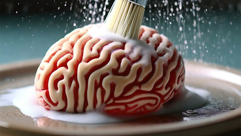
It’s been known for millennia that the human body accumulates waste as a result of day-to-day functioning, but it’s now recognized that the awake, active brain also builds up waste that negatively affects neural function if not removed.
Thankfully, it was recently discovered that the brain has a garbage removal mechanism.
“The glymphatic system is a brain-wide network of perivascular spaces that facilitates the clearance of waste products from the brain during sleep,” Jeffrey Iliff, PhD, professor in the Department of Psychiatry and Behavioral Sciences/Department of Neurology, University of Washington School of Medicine, and researcher at the VA Puget Sound, Seattle, told Medscape Medical News.
This system “serves a similar function in the brain as the lymphatic system does in the rest of the body,” Iliff continued.
As a medical student he learned about the cerebrospinal fluid (CSF) surrounding the brain “we thought of it as an inert fluid with a primarily protective purpose, functioning as a sort of shock absorber,” he said. The reality is far more complex, and it’s now known that the CSF plays “an important part in cleansing brain tissue by removing waste products and conveying nutrients throughout brain tissue.”
The Glymphatic System
When first described in 2012, there was skepticism that a brain waste clearance system existed in humans but a new proof-of-principle study used contrast MRI imaging to visualize the glymphatic system.
Senior author Juan Piantino, MD, MCR, associate professor of pediatrics, Division of Neurology, School of Medicine and physician scientist, Neuroscience Division, Papé Family Research Institute, both in Oregon Health and Science University, Portland, Oregon, said their study was the first to “definitively reveal” the network of perivascular spaces in human beings.
Five patients consented to receiving a gadolinium-based contrast agent through a lumbar drain that was already in place for tumor removal. The tracer was carried via the CSF into the brain and analyzed at different time points following their surgery. The images revealed that fluid moved along defined pathways rather than diffusing uniformly through brain tissue. This finding was documented with fluid attenuated inversion recovery, which showed the perivascular spaces brighten when perfused with the CSF.
Both Iliff and Piantino believe that better understanding the glymphatic system may shed light on the development of neurodegenerative diseases such as Alzheimer’s disease (AD) and may ultimately lead to drugs or other strategies to enhance healthy brain function.
Iliff explained that because there’s no lymphatic circulation within the brain itself, extracellular proteins must be cleared by some alternative mechanism. “[The] CSF might be seen as the ‘sink’ by which extracellular solutes are ‘washed away,’ but it wasn’t clear until relatively recently how these solutes move from the parenchyma to the CSF.”
His research used imaging techniques and fluorescent tracers in a mouse model to demonstrate the drainage pathways.
The glymphatic model suggests that arterial pulsations propel CSF through perivascular spaces formed by the vascular end feet of astrocytes. These spaces, although continuous with the subarachnoid space, also provide “distinct channels” for the rapid flow of CSF into the parenchyma. “The blood vessels in the brain serve as a kind of ‘scaffold’ to organize and support the process,” Iliff said.
CSF flow is enabled through the expression of a water transporter called aquaporin-4 (AQP4). The CSF enters the brain, mixes with the interstitial fluid, and exits via perivenous spaces draining through meningeal and cervical lymphatic vessels or arachnoid granulations. The exiting fluid carries the waste.
Sleep is the most important time during which this “brain housekeeping” takes place, Iliff said. Impairment in this system or in normal sleep can raise the risk for accumulation of undesirable solutes (eg, amyloid-beta), resulting in AD and other neurodegenerative conditions.
What’s in a Name? (and Other Controversies)
The term “glymphatic” was coined to join two ideas: “lymphatic” function which supports waste clearance and “glia” because it’s the brain’s glial cells that support these processes, Iliff recounted.
The term may be new, but there were published reports of a nascent concept. “We collected some of those threads and consolidated them into a single model and connected it to the biology of astrocytes and the biology of sleep” he said. The name ‘glymphatic system’ was coined by Stephen Goldman and Maiken Nedergaard, but the neologism wasn’t well received by all. “Some members of the field felt that by renaming this biology, we were claiming it as our own, and not adequately acknowledging prior research,” added Iliff.
The glymphatic system is connected to a downstream authentic lymphatic network that is associated with the meninges covering the brain as well as cranial nerves and large vessels that exit the skull. Although the anatomical and functional connections between these two networks “aren’t completely understood,” a powerful body of evidence supports the existence of a glymphatic system and its relationship with the meningeal lymphatic network.
Iliff noted additional controversies, adding that “controversy can be a sign that we’re on to something that matters — which is exciting.”
One concerns the presence of AQP4. Its role was demonstrated in multiple lines of research. For example, AQP4 gene deletion exacerbated glymphatic pathway dysfunction after traumatic brain injury (TBI) and promoted the development of neurofibrillary pathology and neurodegeneration. The deletion of AQP4 was found to exacerbate amyloid-beta plaque accumulation in a mouse model of AD. But a 2017 paper called the glymphatic model into question, noting that not all AQP4 data could be replicated. “The discrepancy between findings may lie in differences in experimental methods, rather than being a flaw in understanding the role of APQ4,” Iliff commented.
Another study challenged the notion that a function of sleep is to actively clear metabolites and toxins from the brain. Those researchers used fluorescent tracers to measure clearance and movement of these molecules in the brains of mice and found that movement was actually independent of sleeping, waking, or anesthesia and that brain clearance was markedly reduced, not increased, during sleep and anesthesia.
Iliff believes the study is flawed. “The researchers are essentially quantifying the movement of solutes from one part of the brain to another part of the brain, which we would regard as ‘transport,’ not ‘clearance.’” Additionally, the mice were anesthetized for an hour or so, but studied over many hours, after it had worn off. The authors concept of ‘clearance,’ and ‘sleep,’ “are different from what the rest of the field is doing,” he said.
Sleep, Neurodegeneration, and Clinical Implications
There has been a long-standing clinical association between sleep disruption and dementing disorders such as AD. It was initially assumed that this is due to degeneration in brain regions that regulate sleep. But Iliff pointed to more recent data that suggests that sleep disruption might actually drive the development of the pathology that underlies diseases like AD.
He has reported that the glymphatic system clears amyloid-beta during sleep. More recent data found it clears tau and alpha-synuclein as well.
Conditions that are risk factors for neurodegeneration are also associated with impairment of glymphatic function in animal models, he said, such as obstructive sleep apnea (OSA) — a modifiable risk factor for AD and Parkinson’s disease (PD).
A wide range of neurologic conditions are associated with impaired glymphatic function, including AD, PD, normal pressure hydrocephalus, stroke, cerebral small vessel disease, multiple sclerosis, TBI, migraine, and idiopathic intracranial hypertension. Additional diseases associated with alterations in the glymphatic and meningeal lymphatic system interplay include Huntington disease, frontotemporal dementia, the neuromyelitis optic spectrum disorders, and even brain tumors — particularly gliomas.
There are clinical associations between poor sleep, excessively long or short sleep durations or OSA and dementia. “That, on its own, would suggest paying attention to sleep problems, particularly in midlife, as a potential modifiable risk factor for dementia,” Iliff said.
Piantino agrees. He encourages his patients to “take steps to improve the quality of sleep now, including establishing a relaxing routine and minimizing screens in the bedroom before sleep.”
Other suggestions for keeping your glymphatic system healthy are to improve cardiovascular health since the system relies on arterial pulsatility and physical exercise, which has a positive impact on sleep architecture as well as cardiovascular benefits.
In the future, Iliff envisages being able to identify impairment early using a blood test, imaging scan, or device for example.
He is involved in research on a device described in a recent Nature paper. Healthy older adults wore a head-cap fitted with electrodes to continuously measure sleep-active changes in parenchymal resistance. In two crossover studies participants were subjected to one night of natural sleep and one night of wakefulness, separated by at least 2 weeks. Across both studies, it was found that parenchymal resistance remained constant or increased during periods of waking but declined steadily through periods of sleep.
“We found that the glymphatic system was functioning in deep and REM [rapid eye movement] sleep, and as the person was waking up,” Iliff said. “But the clearance function appeared to accelerate the longer the subject slept and then gradually slow down as the person was waking up.” This was the first time anyone was able to actually track glymphatic function in people at different levels of sleep during a single night.
Applied Cognition, the company that developed the device, is also developing drugs to enhance glymphatic clearance of amyloid and tau in human beings, according to Iliff.
He looks forward to ongoing “exciting” developments, which will pave the way for new therapeutic approaches to the prevention and treatment of neurodegenerative diseases.
Iliff reported receiving research funding from the National Institutes of Health, US Department of Defense, US Department of Veteran’s Affairs, and the Leducq Foundation. He reported serving as the chair for the Scientific Advisory Board for the company Applied Cognition, from which he received compensation and in which he holds equity. Piantino reported receiving research funding from the National Institutes of Health; the National Heart, Lung, and Blood Institute; and the Papé Family Foundation.
Batya Swift Yasgur, MA, LSW is a freelance writer with a counseling practice in Teaneck, New Jersey. She is a regular contributor to numerous medical publications, including Medscape and WebMD, and is the author of several consumer-oriented health books as well as Behind the Burqa: Our Lives in Afghanistan and How We Escaped to Freedom (the memoir of two brave Afghan sisters who told her their story).
