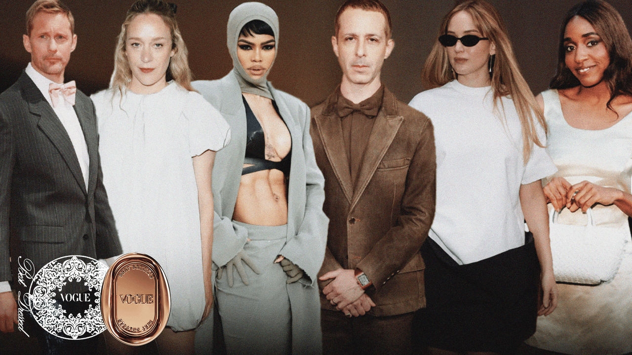
TM9SF1 as a Novel Prognostic Biomarker for Sepsis Severity and Mortali

Introduction

Sepsis is a detrimental organ dysfunctional state that occurs due to the dysregulated host response against infection. This definition was established by the recent International Consensus Definition of Sepsis and Septic Shock (Sepsis-3).1,2 Various microorganisms can cause sepsis,3 with a causative pathogen identified in approximately two-thirds of sepsis patients admitted to intensive care units (ICUs). Both lungs and abdomen are the most common sites of infection.4,5 Globally, around 50 million cases of sepsis occur annually, resulting in approximately 11 million sepsis-related mortalities. The mortality rate associated with sepsis is estimated from 10 to 20% per year.6 In patients with sepsis hospitalized in critical care units, one-third do not survive > 30 days, with fatality rates affected by age, comorbidities, and the number and type of organ malfunctions.7 However, the management of sepsis has remarkably advanced and focused on implementing early and appropriate antimicrobial therapy (with source control where indicated) and comprehensive organ support measures. These strategies have markedly reduced sepsis-related mortality over the past century.8 Despite these developments, the mortality rate associated with sepsis has remained unchanged over the past decade.9 Recent studies have indicated that disease severity and progress predominantly depend on host factors, emphasizing the need to better resolve individual responses at the molecular level.10 Despite multiple genes being associated with sepsis progression, their efficacy in predicting severity and prognosis remains limited (eg, lack of specificity or dynamic range).11–13 Thus, early identification of clinical deterioration continues to pose a significant challenge for clinicians. Therefore, exploring novel biomarkers for early predicting disease status and outcomes in sepsis patients is crucial and urgent.

Sepsis is a complex and heterogeneous clinical status characterized by diverse immunological and physiological responses against infection.14 In 1992, systemic inflammatory response syndrome (SIRS) was described as the systemic inflammatory responses observed in septic patients.15 The excessive inflammatory responses in sepsis are primarily mediated by the activation of the complement and coagulation systems and the involvement of leukocytes, endothelial cells, and cytokines (ie, chemokines and interferons; IFN) secreted by immune cells.16 The unchecked stimulation of proinflammatory cytokines (ie, tumor necrosis factor (TNF) and interleukin-1β (IL-1β) plays a pivotal role in mediating tissue injury, which is further exacerbated by neutrophils stimulation via the secretion of reactive oxygen species (ROSs) proteases, and neutrophil extracellular traps (NETs). These NETs, composed of fiber networks containing proteases, histones, and DNA, contribute significantly to tissue damage.17 The sepsis pathophysiology involves a dysregulated immune response to an invading pathogen, which fails to resolve the homeostasis process. This imbalance leads to a pathological state characterized by prolonged and excessive inflammation, followed by immune suppression. Such mechanisms contribute to the deterioration of the clinical condition in affected patients.18 Molecules involved in the modulation of inflammation may be related to the prognosis of sepsis and have the potential to serve as novel biomarkers for examining the severity and clinical findings of sepsis patients.

Transmembrane 9 superfamily member 1 (TM9SF1) is an autophagy-associated protein,19 initially cloned in 1997.20 Previous studies have depicted that TM9SF1 plays a role in regulating inflammation.21–23 As a member of the TM9SF family, which is defined by an extensive noncytoplasmic region and 9 transmembrane segments, the mRNA level of TM9SF1 is significantly elevated in lung tissue from mice with lipopolysaccharide (LPS)-mediated acute lung injury.23 Furthermore, in TM9SF1 knockout mice, the progression of lung injury was remarkably attenuated, as represented by reduced penetration of inflammatory cells and lowered level inflammatory factors.23 As per the previous findings, which demonstrated a crucial association between TM9SF1 expression, immune regulation, and the advancement of lung inflammatory injury, it can be predicted that changes in TM9SF1 expression may contribute to the onset and progression of sepsis, potentially exacerbating lung injury through the modulation of immune and inflammatory responses.

This study used a prospective cohort design involving patients diagnosed with sepsis who were hospitalized at Xiangyang Central Hospital. Healthy controls (HCs) also participated in this study. The study aimed to examine the differential expression of TM9SF1 in sepsis patients and its potential as a novel biomarker for early disease severity and advancement detection. Further, the predictive models were developed to identify patients at risk for severe sepsis and assess their clinical outcomes.

Materials and Methods

Study Participants

This prospective cohort study was conducted on adult sepsis or septic shock patients who were hospitalized in the respective hospitals’ medical wards or medical ICUs. All study participants (patients and HCs) were recruited between January 1, 2024, and April 30, 2024, with proper follow-up extending until June 30, 2024. The inclusion criteria for patients were being>18 years old and hospitalized due to having similar symptoms like sepsis. The sepsis and septic shock diagnosis criteria were established based on the Third International Consensus Definition for Sepsis and Septic Shock (Sepsis-3): 2016.2 The clinical criteria for septic shock include (1) the requirement for vasopressor treatment to sustain a mean arterial pressure (MAP) of ≥ 65 mmHg and (2) a serum lactate level > 2 mmol/L, which persists despite adequate fluid resuscitation. At the same time, Sequential Organ Failure Assessment (SOFA) score and Acute Physiology and Chronic Health Evaluation (APACHE)-II score were assessed within the first 24 h of admission.2

The study also has exclusion criteria, such as (1) age ≤ 18 years; (2) mortality within 24 h of hospitalization; (3) a history of malignant tumors or hematological disorders; (4) inability to accurately examine the severity of sepsis or septic shock condition; and (5) missing data on age, gender, or key clinical laboratory tests. The primary follow-up endpoints for these patients were either recovery or death, with the follow-up period concluding on the day of discharge of the last patient. Moreover, 64 normal individuals who had undergone routine physical examinations at the hospital and were similar to the sepsis and septic shock patients by gender and age were involved as HCs.

Clinical Information and Sample Collection

Basic clinical data for sepsis and septic shock patients, such as clinical, demographic, and laboratory data, was systematically collected from electronic medical records using standardized forms by expert investigators. Patients were examined from the first day of hospitalization until discharge or death, with the study group remaining non-intrusive and not influencing medical decisions.

Laboratory test analyses within 24 h of hospitalization were evaluated. The following laboratory parameters were critically examined: white blood cell count (WBC), hematocrit (HCT), hemoglobin (Hb), lymphocyte % (LYM%), monocyte % (MON%), neutrophil % (NEU%), neutrophil-lymphocyte ratio (NLR), monocyte-lymphocyte ratio (MLR), platelet-lymphocyte ratio (PLR), platelet count (PLT), platelet distribution width (PDW), mean platelet volume (MPV), procalcitonin (PCT), activated partial thromboplastin time (APTT), prothrombin time (PT), international normalized ratio (INR), thrombin time (TT), D-dimer, erythrocyte sedimentation rate (ESR), C-reactive protein (CRP). Oxygenation index (OI) was calculated as the PaO2/FiO2 ratio in sepsis patients.

These parameters were analyzed using an Atellica Solution Immunoassay and Clinical Chemistry Analyzer (Siemens Healthcare Diagnostics, Erlangen, Germany). Both WBC and LYM % were determined using the Sysmex XE-2100 hematology analyzer (TOA Medical Electronics, Kobe, Japan). APTT, PT, INR, fibrinogen, and D-dimer levels were analyzed with the Sysmex CS-5100 System (Siemens Healthcare Diagnostics, Erlangen, Germany). CRP levels were quantified using the i-CHROMA device (BodiTech Med Inc., Chuncheon, Korea). ESR was measured using the Vacuette ESR System (Greiner Bio-One GmbH, Frickenhausen, Germany).

Blood samples were collected during hospitalization during the routine procedure of complete blood count after admission to the clinical ward or ICU. Blood samples from 45 sepsis and septic shock patients were collected at the time of admission and the end of the follow-up period.

RNA Extraction and Analysis of TM9SF1 and Cytokine Expression via RT-qPCR

The peripheral blood mononuclear cells (PBMCs) were isolated from 2 mL of residual blood following a density gradient centrifugation method. Specifically, whole blood samples were subjected to centrifugation at 3000 rpm for 20 minutes using Human Lymphocyte Separation Medium (Solarbio, P8610). The PBMC layer was collected and resuspended in TRIzol reagent (Invitrogen, 15596026) before storage at −80°C.

For RNA extraction, TRIzol-suspended PBMC samples were thawed on ice. Subsequently, 200 μL of chloroform was added, followed by thorough mixing and incubation on ice for 15 minutes. After centrifugation at 12,000 rpm for 15 minutes at 4°C, the aqueous phase was carefully transferred to a new tube to avoid the interphase. An equal volume of isopropanol was added, mixed, and incubated on ice for 15 minutes. The mixture was then subjected to centrifugation under the same conditions. The resulting RNA pellet was washed with 75% ethanol, air – dried, and dissolved in RNase – free water.

The extracted RNA was used for reverse transcription-quantitative PCR (RT-qPCR) analysis to measure the expression levels of TM9SF1 as well as various cytokines including FOXP3, TNF-α, IFN-γ, IL −17A, and IL-6. For this purpose, the RNA samples were reverse transcribed into cDNA using a reverse transcription kit (iScript cDNA synthesis kit, Bio-Rad, 1708891). qPCR was performed in a reaction mixture containing cDNA, specific primers (listed in Table S1), and a qPCR master mix (iTaq Universal SYBR Green Supermix and Bio-Rad, 1725125) on a qPCR instrument (ABI7500). The thermal cycling conditions were as follows: initial denaturation at 95°C for 5 minutes, followed by 40 cycles of denaturation at 95°C for 15 seconds and annealing/extension at 60°C for 30 seconds. The relative expression levels of the target cytokines were calculated using the 2^−ΔΔCt method, with the GAPDH serving as the internal reference.22

Statistical Analysis

Continuous variables were represented as mean ± standard deviation or median (interquartile range), while qualitative data was depicted as N with corresponding percentages. The Student’s t-test was used to compare differences in variables for quantitative analysis, whereas the chi-squared test was applied to assess differences in qualitative analysis. Spearman correlation analysis examined the correlation between TM9SF1 mRNA expression and biochemical indices. Univariate and multivariate logistic regression analyses were used to determine multivariable-adjusted odds ratios (ORs) with 95% confidence intervals (CIs) for the correlation between TM9SF1 level and sepsis severity. The combined analyses of disease advancement and mortality were evaluated via Cox regression. Both hazards ratios (HRs) and their 95% CIs were also determined. Covariates included in the adjustments were age, gender, status of smoking, drinking, and diseases history.

Receiver operating characteristic (ROC) curve analysis evaluated the predictive accuracy of TM9SF1, the key indicator, and the prediction model for sepsis severity and mortality. Prediction models were developed for sepsis severity and mortality by selecting variables (p < 0.05) from univariate analysis with a combination of multivariate regression analysis. After adjusting for other covariates, the final prediction model included variables with p < 0.05. As per the results of the multivariate analysis, a nomogram was developed to predict sepsis severity or mortality. The nomogram’s performance was validated by discrimination (a corrected Harrell’s concordance index [C-index]) and calibration (calibration curves and the Hosmer–Lemeshow test), using bootstrapping with 1000 resamples. Kaplan-Meier survival curve analysis was performed to analyze the risk of mortality.

PASS 2021 software was used to calculate the minimum sample size. The parameters were set as follows: significance level (α=0.05, two-sided), statistical power [(1-β)=0.90], expected effect size (OR = 2.0, HR = 2.0), and the follow-up dropout rate of the participants (10%) was also considered. A two-sided p < 0.05 was regarded as statistical significance. Graphs were plotted via GraphPad Prism 5 and R v3.6.3 software, and statistical analyses were conducted via SAS software v9.4 (SAS Institute, Cary, NC).

Results

Clinical and Demographic Features of the Study Population

From January 1, 2024, and April 30, 2024, approximately 225 adult sepsis patients were hospitalized at Xiangyang Central Hospital. Among them, 118 patients were identified as having septic shock, while 107 were classified as having sepsis. The septic shock cohort’s demographic features are depicted in Table 1. No significant difference in average age was observed between both groups (66.52 ± 17.03 vs 70.36 ± 14.38, p = 0.068). However, the septic shock group had a considerably higher male distribution than the sepsis group (male: 58.9% vs 72.9%, p = 0.027). Among sepsis patients, 67.9% had one comorbidity, and it was more prevalent in the septic shock than in the sepsis patients (Table 1). The mean average of SOFA score for the overall patients was 9.68 ± 2.36, and was markedly higher in the septic shock group relative to the sepsis group (11.53 ± 2.64 vs 7.92 ± 1.47, p < 0.001), reflecting a more severe clinical condition, as well as APACHE-II score (22.56 ± 5.73 vs 19.72 ± 4.96, p < 0.001).

Table 1 Demographic Characteristics of Patients with Sepsis (Sepsis vs Septic Shock Group)

All laboratory findings, stratified by disease severity, are outlined in Table 2. In the blood routine examination, patients with septic shock showed substantially higher CRP and ESR (all p < 0.001) but lower Hb, LYM%, and MON% than those with sepsis (all p < 0.05). In addition, significant variances were observed in WBC, NEU%, neutrophils, lymphocytes, NLR, PLR, and MLR (all p < 0.05). In the case of coagulation markers, the septic shock group demonstrated elevated levels of INR, TT, and D-dimer (all p < 0.05). Furthermore, the average OI was lower in the septic shock group than in sepsis group (p < 0.001). The level of PaO2 and lactate was also significantly different between the septic shock patients and the sepsis patients (p = 0.030 and p < 0.001).

Table 2 Laboratory Parameters of Patients with Sepsis (Sepsis vs Septic Shock Group)

The study comprised all sepsis patients who completed their follow-up until June 30, 2024. Among them, 78 (34.7%) died, and 147 (65.3%) progressed during their follow-up period. Patients who died and those who progressed from septic shock showed similar outcomes regarding their demographic and clinical features. Non-survivor patients depicted worse laboratory parameters compared to those who survived, such as higher WBC, NEU%, PLR, NLR, MLR, ESR, CRP, D-dimer, SOFA score, APACHE-II score, and lactate, as well as lower LYM%, MON%, PaO2, SaO2, and OI (Tables 3 and 4).

Table 3 Demographic Characteristics of Patients with Septic Shock (Survivor vs Non-Survivor)

Table 4 Laboratory Parameters of Patients with Septic Shock (Survivor vs Non-Survivor)

Upregulation of TM9SF1 in Severe Sepsis Patients

The differential expression of TM9SF1 was assessed in all groups via qPCR, as illustrated in Figure 1. Blood samples were collected from 45 sepsis patients during admission and the follow-up time points. As shown in Figure 1A, TM9SF1 mRNA level was substantially higher in sepsis patients relative to HCs (sepsis patients: 0.25 ± 0.02; HCs: 0.10 ± 0.02; p < 0.001). The variations in TM9SF1 expressions between septic shock group and sepsis group were also examined (Figure 1B). Its mRNA expression was markedly higher in septic shock patients than in sepsis patients (0.29 ± 0.02 vs 0.16 ± 0.01, p < 0.001). A paired t-test compared TM9SF1 expression between the severe and non-severe patients’ states (Figure 1C). The findings revealed that as the condition of sepsis patients improved and changed from severe to non-severe, the mRNA levels of TM9SF1 decreased considerably (0.25 ± 0.04 vs 0.13 ± 0.02, p < 0.001). Further, to evaluate the disparity in TM9SF1 expression between patients who had progressed and those who died, independent-sample t-tests were conducted (Figure 1D and E). The findings suggested that the mRNA level of TM9SF1 elevated substantially in non-survivor patients than that in survivors, both in all the sepsis patients (0.30 ± 0.03 vs 0.19 ± 0.02, p < 0.001) and septic shock patients (0.37 ± 0.04 vs 0.23 ± 0.02, p = 0.005).

Figure 1 Upregulation of TM9SF1 in patients with severe sepsis. (A) Patients with sepsis and HC group. (B) Patients with sepsis and septic shock conditions. (C) Paired samples of patients with sepsis, and septic shock group. (D) Patients with sepsis, survival, and non-survival groups. (E) Patients with septic shock, survival, and non-survival groups. **p < 0.01; ***p < 0.001.

TM9SF1 Expression Correlates with Cytokines and Clinical Parameters in Sepsis

Abundant cytokine production is a hallmark of sepsis and is a key contributor to tissue damage associated with this condition.24 Spearman correlation analyses were conducted to investigate the correlation between TM9SF1 mRNA and cytokine levels in patients with sepsis. These analyses were carried out in all sepsis patients, revealed substantial positive relationship between the expression of TM9SF1 and several key cytokines, including IFN-γ (r = 0.788, p < 0.001), TNF-α (r = 0.610, p < 0.001), IL-6 (r = 0.646, p < 0.001), IL-17A (r = 0.818, p < 0.001), and forkhead box P3 (FOXP3) (r = 0.607, p < 0.001) (Figure 2). Furthermore, a subgroup analysis focusing on patients with septic shock demonstrated that the strong associations persisted, particularly for IFN-γ (r = 0.834, p < 0.001), IL-6 (r = 0.784, p < 0.001), TNF-α (r = 0.709, p < 0.001). These findings indicate a robust correlation between plasma cytokine levels and TM9SF1 expression, illustrating that it may play a regulatory role in the harsh microenvironment of sepsis. Moreover, correlation analyses examining the association between each cytokines, as well as TM9SF1 expression in relation to clinical parameters are illustrated in Figure S1 and Table S2.

Figure 2 The significant correlation of TM9SF1 expression with cytokines in patients with sepsis. (A) FOXP3. (B) TNF-α. (C) IFN-γ. (D) IL-17A. (E) IL-6. FOXP3, forkhead box P3; TNF-α, tumor necrosis factor-α; IFN-γ, interferon-γ; IL-17A, interleukin-17A; IL-6, interleukin-6.

Abbreviation: CI, confidence interval.

Upregulation of TM9SF1 Enhances the Severity and Mortality of Sepsis

Univariable and multivariable logistic regression analyses assessed the correlation between TM9SF1 level and sepsis severity (Table S3). After adjusting for age, gender, smoking status, drinking status, and diseases history, higher levels of TM9SF1 were considerably correlated with increased sepsis severity (OR = 47.14, 95% CI = 8.06–275.65, p < 0.001). When patients were stratified into high- and low-risk groups as per the median TM9SF1 level (0.14), those in the high-risk group demonstrated a 2.94-fold greater likelihood of severe sepsis relative to the low-risk group (OR = 3.29, 95% CI = 1.88–5.78, p < 0.001).

To further investigate the correlation between TM9SF1 expression and patient mortality, Cox regression analysis and Kaplan-Meier survival curve were carried out (Table S4 and Figure S2). Patient mortality increased as the level of TM9SF1 increased, adjusting for age, gender, smoking status, drinking status, and diseases history (HR = 37.21, 95% CI = 12.80–108.21, p < 0.001). Moreover, patients with elevated TM9SF1 expression had shorter survival times than those with lower levels (HR = 11.12, 95% CI = 4.35–28.45, p < 0.001).

Higher Predictive Potential of TM9SF1 Over Clinical Indicators for Sepsis Severity and Mortality

ROC curve analysis estimated the predictive abilities of TM9SF1 and various clinical indicators for the severity and mortality of sepsis. Table 5 shows the efficacy of predicting sepsis severity using TM9SF1 and several clinical indicators, including WBC, LYM %, MON%, NEU%, NLR, PLR, MLR, CRP, ESR, D-dimer, SaO2, OI, and lactate. Among all the independent variables, TM9SF1 possessed the highest area under the ROC curve (AUC = 0.822, 95% CI = 0.766–0.878). The AUC for severity prediction was 0.768 (95% CI = 0.705–0.832) for ESR and 0.615 (95% CI = 0.541–0.688) for OI. TM9SF1 was the best marker to distinguish between sepsis and septic shock patients. A cut-off value of 0.10 for the TM9SF1 expression level showed a sensitivity of 89.4% and a specificity of 72.9%.

Table 5 The Predictive Ability of Prediction Nomogram and Various Clinical Indicators for the Severity of Sepsis

The predictive potential of TM9SF1 for patient mortality was evaluated and compared with other independent variables using time-dependent ROC analysis (Table 6). TM9SF1 demonstrated higher predictive accuracy, achieving a Harrell’s C-index of 0.853 (95% CI = 0.782–0.924), with a sensitivity 91.8%, specificity 78.3%, and a cut-off value of 0.30. In comparison, the Harrell’s C-index for mortality prediction was 0.680 (95% CI = 0.584–0.775) for ESR, 0.668 (95% CI = 0.567–0.769) for NLR, and 0.749 (95% CI = 0.654–0.874) for lactate. These findings highlight the higher predictive capability of TM9SF1 compared to several clinical indicators for assessing sepsis’s severity and mortality risk. Thus, TM9SF1 may serve as a promising novel biomarker for predicting sepsis outcomes.

Table 6 The Predictive Ability of Prediction Nomogram and Various Clinical Indicators for the Mortality of Sepsis with Time-Dependent ROC Analysis

Establishment and Validation of the Nomogram

To develop a prediction model for the severity of sepsis, multivariate logistic regression analysis was carried out with variables having p values < 0.05 in univariate logistic regression analysis. The final prediction model included variables with p < 0.05 after adjusting for other covariates. As a result, TM9SF1, D-dimer, ESR and CRP were the independent risk factors that predicted sepsis severity, with ORs of 47.81 (95% CI = 28.33–74.41, p < 0.001), 1.20 (95% CI = 1.10–1.30, p < 0.001), 1.04 (95% CI = 1.02–1.05, p < 0.001), and 1.13 (95% CI = 1.02–1.47, p = 0.040), respectively.

The mortality rate of sepsis patients was analyzed using multivariable Cox proportional hazards regression analysis. The findings revealed that the mortality risk of septic shock patients was independently correlated with a higher TM9SF1 level (HR = 28.69, 95% CI = 17.19–47.48, p < 0.001), CRP (HR = 1.14, 95% CI = 1.03–3.07, p = 0.027), ESR (HR = 1.05, 95% CI = 1.01–1,37, p = 0.046), lactate (HR = 8.69, 95% CI = 5.01–11.13, p < 0.001), and OI (HR = 0.08, 95% CI = 0.04–0.26, p = 0.047).

To provide a quantitative tool capable of predicting the individual probability of developing septic shock, a novel nomogram incorporating TM9SF1, D-dimer, ESR, and CRP level was constructed in line with the multivariable logistic analysis (Figure 3). The probability of developing septic shock was calculated for each patient by adding the scores of the aforementioned four variables. Likewise, a prognostic nomogram for mortality prediction of patients with septic shock was created based on the significant independent predictors listed in Figure 4, namely, TM9SF1, ESR, CRP, lactate, and OI. The mortality probability of the patients was calculated by adding the predictors included in the nomogram. The nomogram developed for predicting sepsis severity was validated using the ROC curve, demonstrating an AUC of 0.883 (95% CI = 0.839–0.927) with a sensitivity 91.5% and a specificity 78.4% (Table 5). For mortality prediction in patients with septic shock, the nomogram achieved a C-index of 0.931 (95% CI: 0.884–0.978, Table 6).

Figure 3 Severity prediction nomogram incorporating TM9SF1 and inflammatory markers.

Figure 4 Prediction nomogram for the mortality of sepsis.

Abbreviation: CRP, C-reactive protein; ESR, erythrocyte sedimentation rate; OI, oxygenation index.

In addition, patients were stratified into high- and low-risk groups as per the total score to aid prognostic discrimination. The optimal cut-off value for total points was 44.30 for effective risk classification. Kaplan-Meier survival curve analysis was performed to analyze the risk of death in patients with sepsis shock at different points calculated by the prognostic nomogram (Figure 5). It was obvious that when patients were stratified into high- and low-risk groups, those in the high-risk group demonstrated a 8.39-fold greater likelihood of severe sepsis relative to the low-risk group (HR = 8.39, 95% CI = 3.74–18.80, p < 0.001).

Figure 5 Kaplan-Meier survival curve analysis in patients with sepsis shock. The curve is stratified by the cut-off value for total points (44.3) of prognostic nomogram in patients with sepsis shock.

Abbreviation: HR, hazard ratio.

Discussion

This study first time evaluated the role of TM9SF1 in the early identification of disease severity and mortality in sepsis or septic shock patients. TM9SF1 mRNA level was markedly observed in septic shock patients relative to those with sepsis group. Further, multiple logistic regression analysis demonstrated a positive correlation between disease severity and TM9SF1 levels. Moreover, the Cox regression analysis indicated that elevated TM9SF1 expression was associated with high mortality in septic shock patients. ROC curve analysis demonstrated that TM9SF1 outperformed several clinical markers in examining both the severity and mortality of sepsis. The current prediction models showed strong efficacy in identifying disease severity and early prognosis. Moreover, TM9SF1 mRNA expression was strongly correlated with the levels of cytokines and various clinical parameters. These results depict that TM9SF1 may be served as a potential novel biomarker, provide individualized intervention and treatment for high-risk patients of sepsis in advance through the severity and mortality prediction model. Meanwhile, TM9SF1 may have important reference value in the targeted therapy of sepsis.

Despite its higher costs, qPCR offers significant advantages for critical sepsis cases: non-invasive sample accessibility (using PBMCs rather than invasive procedures like bronchoalveolar lavage or pleural/ascitic fluid sampling) and a short in-laboratory turnaround time. These strengths enable timely therapeutic guidance, which is strongly aligned with the 2021 Surviving Sepsis Campaign Guidelines’ mandate for early recognition and resuscitation. Consequently, the clinical value of qPCR for specific biomarkers significantly outweighs its costs. Furthermore, patients in pulmonary and hematology departments exhibiting elevated TM9SF1 expression might benefit from earlier transfer to the ICU.

According to reports, sepsis is related to numerous biomarkers and clinical parameters.11–13 Several studies have investigated the use of NLR, PLR, or MLR as indicators of clinical course for patients suffering from sepsis, which had significant prognostic value in predicting the mortality of sepsis patients.25,26 Moreover, a retrospective cohort study showed that several clinical variables could be considered dependent risk factors for predicting and assessing severity in sepsis patients, such as age, gender, PaO2/FiO2 ratio, and SOFA score.27 The mRNA expression pattern in PBMCs also demonstrated substantial potential for clinical application in sepsis. Elevated mRNA levels of CD177 and MMP8 in PBMCs were highly valuable for diagnosing patients with septic shock.28 Autophagy-associated genes, including GABARAPL2 and GAPDH, have been recognized as key hub genes in 7 differentially infiltrated immune cell types and hold potential as diagnostic markers for sepsis.29

The excessive production of inflammatory mediators released by neutrophils and macrophages during the early stages of sepsis is closely related to disease severity and prognosis.30 Both IL-6 and TNF-α, which are secreted by activated monocytes, serve as prognostic risk factors for deaths in sepsis patients.31 Single-cell transcriptomic analysis of PBMCs from sepsis patients revealed a remarkable enrichment of genes associated with the IL-17 signaling pathway in monocytes, NK cells, and T cells.32 This study confirmed the association between TM9SF1 expression and these cytokines, which implied that TM9SF1 may be involved in regulating the activation and proliferation of various immune cells in PBMCs. Previous research indicated that the levels of TM9SF1 were elevated in some inflammatory diseases,21–23 and knock-out of TM9SF1 alleviated the symptoms in animal models.21,23 TM9SF1, an intracellular transmembrane protein, possibly regulates the activity of the inflammatory factor signaling pathway, thereby contributing to both the development and progression of inflammation. The inflammatory damage in sepsis patients is expected to be reduced by exploring drugs that target the TM9SF1 molecule. Single-cell RNA sequences may reveal the impact of upregulated TM9SF1 expression on various immune cells; however, this technology would be relatively expensive and time-consuming for clinical application. To investigate whether TM9SF1 modulates the inflammatory cytokine signaling pathway, further in vitro experiments need to be conducted in each immune cell.

Sepsis is a complex immune-inflammatory response that occurs due to infection, and patients can present with varying degrees of immune dysregulation, such as hyperinflammation.33 The results showed that NEU count was substantially upregulated in SSG compared to SG. As a rapid and low-specificity response,34 cytokine storms induced by IL-1 originating from macrophages may result in shock, multiple organ dysfunction such as ARDS, liver dysfunction, and disseminated intravascular coagulation (DIC).35 Anti-infective agents are advised to be administered within one hour following a sepsis diagnosis, as delaying antibiotic treatment elevates the mortality rate among sepsis patients and enhances the risk of progressing to severe sepsis or septic shock.36 However, cytokine levels in clinical laboratories and other clinical indicators remain insufficient to determine the timing of initial medication administration.37 In this study, predictive models based on TM9SF1 were constructed and further combined with both clinical indicators and epigenetic biomarkers, thereby considering the interactions of these factors comprehensively. Both prediction models showed satisfactory prediction efficacy better than that of each clinical indicator alone.

Sepsis may also modify energy metabolism in neutrophils and other immune cells,38 necessitating more energy to maintain antimicrobial functions. TM9SF1 may also participate in the energy metabolism of cells that regulate both adaptive and innate immune responses. TM9SF1 was first recognized as a marker correlated with autophagy and has been implicated in both autophagy19 and the modulation of inflammatory variables. Increased levels of TM9SF1 have been correlated with the severity of acute lung injury, as shown in our previous results.23 Autophagy is initiated to eliminate unwanted components, including bacteria and malfunctioning mitochondria,39 in response to a state of stress. The recycling of cellular products and the maintenance of energy supply are essential for the elimination of invading pathogens by immune cells in a highly active state.40 While hyper-inflammation is favorable during the early stage of sepsis, T cell exhaustion may predispose patients to future infections due to sepsis-induced immunosuppression, especially in elderly patients.41 The study reported that TM9SF1 plays a crucial role in inflammatory autoimmune disease by regulating autophagy.21 Research indicated that autophagy rises early in sepsis-induced acute kidney injury (AKI) and falls afterward in animal models, one study found that markers of autophagy increased at 3h after establishing sepsis-induced AKI.42 These results are consistent with the trend of TM9SF1 expression level in sepsis patients and support the idea that TM9SF1 can predict organ damage and the severity of sepsis in its early stages.

This study was the first to investigate the critical role of TM9SF1 expression in the early identification of disease severity and mortality among sepsis patients. As per the large population and various disease types, the study provides novel evidence of a substantial association between TM9SF1 expression and sepsis progression. As visualized through nomograms, the innovative prediction models based on TM9SF1 expression demonstrated strong efficacy in analyzing disease status and clinical outcomes in sepsis patients. However, the study has certain limitations. Firstly, all patients included in this study were recruited from Xiangyang Central Hospital. Expanding the cohort to include participants from various regions across the country is recommended to validate the scientific accuracy and representativeness of the findings. A multicenter study with longer follow-up has been planned to strengthen the reliability of the conclusions. Secondly, no direct mechanistic link between TM9SF1 and sepsis pathogenesis has been established yet. TM9SF1 may participate in the pathogenesis and progression of sepsis by stimulating production of inflammatory cytokines in various immune cells or participating in the activation and/or maturation of immune cells. Its involvement in sepsis pathogenesis can be investigated by observing alterations in various immune cell activity through TM9SF1 knockout in animal models. For example, animal sepsis models (myeloid-specific TM9SF1 knockout) could be used to analyze whether TM9SF1 actively drives pathogenesis via cytokine dysregulation or merely reflects inflammatory burden.

Conclusion

This study highlights that TM9SF1 mRNA level is substantially upregulated in patients with severe sepsis, and its levels are correlated with higher disease severity and mortality. Its predictive capability for sepsis severity and prognosis surpasses several traditional clinical indicators. Prediction models based on TM9SF1 demonstrated satisfactory performance in the early diagnosis of disease severity and clinical findings. These results underscore the potential of TM9SF1 as a novel indicator for the early assessment and management of sepsis, offering a valuable tool for enhancing patient care and guiding therapeutic strategies.

Data Sharing Statement

The original contributions presented in the study are included in the article/Supplementary Material. Further inquiries can be directed to the corresponding author.

Ethical Statement

The study was conducted in accordance with the Declaration of Helsinki (as revised in 2013). The study protocol was approved by the Ethics Committee of Hubei University of Arts and Science. Informed written consent was obtained from all subject participants in the study. The authors are accountable for all aspects of the work, including ensuring that any questions related to the accuracy or integrity of any part of the work were appropriately investigated and resolved.

Acknowledgments

The authors would like to thank all the reviewers who participated in the review and MJEditor (www.mjeditor.com) for its linguistic assistance during the preparation of this manuscript.

Author Contributions

All authors made a significant contribution to the work reported, whether that is in the conception, study design, execution, acquisition of data, analysis and interpretation, or in all these areas; took part in drafting, revising or critically reviewing the article; gave final approval of the version to be published; have agreed on the journal to which the article has been submitted; and agree to be accountable for all aspects of the work.

Funding

This work was supported by the Hubei Provincial Natural Science Foundation of China (2022CFD109 and 2024AFB165), the Hubei Provincial Natural Science Foundation Innovation and Development Joint Fund Project of China (2025AFD034) and Xiangyang Medical-health Areas Science and Technology Program (2021YL047).

Disclosure

The authors declare no conflicts of interest in this work.

References

1. Rhee C, Zhang Z, Kadri SS, et al. Sepsis surveillance using adult sepsis events simplified eSOFA criteria versus sepsis-3 sequential organ failure assessment criteria. Crit Care Med. 2019;47(3):307–314. doi:10.1097/CCM.0000000000003521

2. Singer M, Deutschman CS, Seymour CW, et al. The third international consensus definitions for sepsis and septic shock (Sepsis-3). JAMA. 2016;315(8):801–810. doi:10.1001/jama.2016.0287

3. Esposito S, De Simone G, Boccia G, De Caro F, Pagliano P. Sepsis and septic shock: new definitions, new diagnostic and therapeutic approaches. J Global Antimicrob Resist. 2017;10:204–212. doi:10.1016/j.jgar.2017.06.013

4. Gauer R, Forbes D, Boyer N. Sepsis: diagnosis and management. Am Family Phys. 2020;101(7):409–418.

5. Vucelic V, Klobucar I, Duras-Cuculic B, et al. Sepsis and septic shock – an observational study of the incidence, management, and mortality predictors in a medical intensive care unit. Croatian Med J. 2020;61(5):429–439. doi:10.3325/cmj.2020.61.429

6. Chiu C, Legrand M. Epidemiology of sepsis and septic shock. Curr Opinion Anaesthesiol. 2021;34(2):71–76. doi:10.1097/ACO.0000000000000958

7. Gu X, Zhou F, Wang Y, Fan G, Cao B. Respiratory viral sepsis: epidemiology, pathophysiology, diagnosis and treatment. Eur Respir Rev. 2020;29(157):200038. doi:10.1183/16000617.0038-2020

8. Jarczak D, Kluge S, Nierhaus A. Sepsis-pathophysiology and therapeutic concepts. Front Med. 2021;8:628302. doi:10.3389/fmed.2021.628302

9. Markwart R, Saito H, Harder T, et al. Epidemiology and burden of sepsis acquired in hospitals and intensive care units: a systematic review and meta-analysis. Intensive Care Med. 2020;46(8):1536–1551. doi:10.1007/s00134-020-06106-2

10. Guarino M, Perna B, Cesaro AE, et al. Update on sepsis and septic shock in adult patients: management in the emergency department. J Clin Med. 2023;12(9):3188. doi:10.3390/jcm12093188

11. Saxena J, Das S, Kumar A, et al. Biomarkers in sepsis. Int J Clin Chem. 2024;562:119891. doi:10.1016/j.cca.2024.119891

12. Yagmur E, Sckaer JH, Koek GH, et al. Elevated MR-proANP plasma concentrations are associated with sepsis and predict mortality in critically ill patients. J Transl Med. 2019;17(1):415. doi:10.1186/s12967-019-02165-2

13. Zhang D, Xu C, Zhang J, et al. Plasma TNFRSF11B as a new predictive inflammatory marker of sepsis-ARDS with endothelial dysfunction. J Proteome Res. 2023;22(11):3640–3651. doi:10.1021/acs.jproteome.3c00576

14. Wang W, Liu CF. Sepsis heterogeneity. World J Pediatrics. 2023;19(10):919–927. doi:10.1007/s12519-023-00689-8

15. American College of Chest Physicians/Society of Critical Care Medicine Consensus Conference. Definitions for sepsis and organ failure and guidelines for the use of innovative therapies in sepsis. Crit Care Med. 1992;20(6):864–874. doi:10.1097/00003246-199206000-00025

16. van der Poll T, Shankar-Hari M, Wiersinga WJ. The immunology of sepsis. Immunity. 2021;54(11):2450–2464. doi:10.1016/j.immuni.2021.10.012

17. McDonald B, Davis RP, Kim S-J, et al. Platelets and neutrophil extracellular traps collaborate to promote intravascular coagulation during sepsis in mice. Blood. 2017;129(10):1357–1367. doi:10.1182/blood-2016-09-741298

18. Cao M, Wang G, Xie J. Immune dysregulation in sepsis: experiences, lessons and perspectives. Cell Death Discovery. 2023;9(1):465. doi:10.1038/s41420-023-01766-7

19. He P, Peng Z, Luo Y, et al. High-throughput functional screening for autophagy-related genes and identification of TM9SF1 as an autophagosome-inducing gene. Autophagy. 2009;5(1):52–60. doi:10.4161/auto.5.1.7247

20. Chluba-de Tapia J, de Tapia M, Jaggin V, Eberle AN. Cloning of a human multispanning membrane protein cDNA: evidence for a new protein family. Gene. 1997;197(1–2):195–204. doi:10.1016/S0378-1119(97)00263-1

21. Xiao J, Zhao Z, Zhou F, et al. TM9SF1 expression correlates with autoimmune disease activity and regulates antibody production through mTOR-dependent autophagy. BMC Med. 2024;22(1):502. doi:10.1186/s12916-024-03729-w

22. Cao F, Zhang L, Zhao Z, et al. TM9SF1 offers utility as an efficient predictor of clinical severity and mortality among acute respiratory distress syndrome patients. Front Immunol. 2024;15:1408406. doi:10.3389/fimmu.2024.1408406

23. Xiao J, Shen X, Chen H, et al. TM9SF1 knockdown decreases inflammation by enhancing autophagy in a mouse model of acute lung injury. Heliyon. 2022;8(12):e12092. doi:10.1016/j.heliyon.2022.e12092

24. Carcillo JA, Shakoory B. Cytokine storm and sepsis-induced multiple organ dysfunction syndrome. Adv Exp Med Biol. 2024;1448:441–457.

25. Zhang Y, Peng W, Zheng X. The prognostic value of the combined neutrophil-to-lymphocyte ratio (NLR) and neutrophil-to-platelet ratio (NPR) in sepsis. Sci Rep. 2024;14(1):15075. doi:10.1038/s41598-024-64469-8

26. Liang P, Yu F. Value of CRP, PCT, and NLR in prediction of severity and prognosis of patients with bloodstream infections and sepsis. Front Surg. 2022;9:857218. doi:10.3389/fsurg.2022.857218

27. Li T, Hu WQ, Li X, et al. Prognostic value of PaO(2)/FiO(2), SOFA and D-dimer in elderly patients with sepsis. J Int Med Res. 2022;50(6):3000605221100755. doi:10.1177/03000605221100755

28. Kong C, Zhu Y, Xie X, Wu J, Qian M. Six potential biomarkers in septic shock: a deep bioinformatics and prospective observational study. Front Immunol. 2023;14:1184700. doi:10.3389/fimmu.2023.1184700

29. Di C, Du Y, Zhang R, Zhang L, Wang S. Identification of autophagy-related genes and immune cell infiltration characteristics in sepsis via bioinformatic analysis. J Thoracic Dis. 2023;15(4):1770–1784. doi:10.21037/jtd-23-312

30. Chousterman BG, Swirski FK, Weber GF. Cytokine storm and sepsis disease pathogenesis. Semin Immunopathol. 2017;39(5):517–528. doi:10.1007/s00281-017-0639-8

31. Li J, Xiao C, Zheng H. Prognostic value of inflammatory cytokine detection for sepsis patients in ICU: a meta-analysis. Am J Transl Res. 2024;16(6):2612–2621. doi:10.62347/NYLM7723

32. Zhao H, Li Y, Sun G, Cheng M, Ding X, Wang K. Single-cell transcriptional gene signature analysis identifies IL-17 signaling pathway as the key pathway in sepsis. Immunobiology. 2023;228(6):152763. doi:10.1016/j.imbio.2023.152763

33. Li L, Peng X, Guo L, Zhao Y, Cheng Q. Sepsis causes heart injury through endoplasmic reticulum stress-mediated apoptosis signaling pathway. Int J Clin Exp Pathol. 2020;13(5):964–971.

34. Doganyigit Z, Eroglu E, Akyuz E. Inflammatory mediators of cytokines and chemokines in sepsis: from bench to bedside. Hum Exp Toxicol. 2022;41:9603271221078871. doi:10.1177/09603271221078871

35. Shakoory B, Carcillo JA, Chatham WW, et al. Interleukin-1 receptor blockade is associated with reduced mortality in sepsis patients with features of macrophage activation syndrome: reanalysis of a prior phase III trial. Crit Care Med. 2016;44(2):275–281. doi:10.1097/CCM.0000000000001402

36. Liu VX, Fielding-Singh V, Greene JD, et al. The timing of early antibiotics and hospital mortality in sepsis. Am J Respir Crit Care Med. 2017;196(7):856–863. doi:10.1164/rccm.201609-1848OC

37. Mitzner S, Kogelmann K, Ince C, Molnar Z, Ferrer R, Nierhaus A. Adjunctive hemoadsorption therapy with cytosorb in patients with septic/vasoplegic shock: a best practice consensus statement. J Clin Med. 2023;12(23):7199. doi:10.3390/jcm12237199

38. Pan T, Sun S, Chen Y, et al. Immune effects of PI3K/Akt/HIF-1alpha-regulated glycolysis in polymorphonuclear neutrophils during sepsis. Critical Care. 2022;26(1):29. doi:10.1186/s13054-022-03893-6

39. Yamashita SI, Kanki T. How autophagy eats large mitochondria: autophagosome formation coupled with mitochondrial fragmentation. Autophagy. 2017;13(5):980–981. doi:10.1080/15548627.2017.1291113

40. Cao C, Yu M, Chai Y. Pathological alteration and therapeutic implications of sepsis-induced immune cell apoptosis. Cell Death Dis. 2019;10(10):782. doi:10.1038/s41419-019-2015-1

41. Inoue S, Suzuki K, Komori Y, et al. Persistent inflammation and T cell exhaustion in severe sepsis in the elderly. Critical Care. 2014;18(3):R130. doi:10.1186/cc13941

42. Zhao S, Liao J, Shen M, Li X, Wu M. Epigenetic dysregulation of autophagy in sepsis-induced acute kidney injury: the underlying mechanisms for renoprotection. Front Immunol. 2023;14:1180866. doi:10.3389/fimmu.2023.1180866

Continue Reading