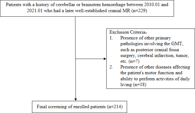
Hypertrophic Olivary Degeneration (HOD) is a form of transsynaptic degeneration caused by lesions in the dentato-rubro-olivary pathway. It was first described by Oppenheim in 1887 during autopsy. In 1931, Guillain and Mollaret proposed that the onset of HOD was associated with damage to the dentato-rubro-olivary pathway and detailed its anatomical relationship. The dentate nucleus connects to the contralateral red nucleus through the superior cerebellar peduncle and the dentato-rubral tract, while the red nucleus connects to the ipsilateral ION via the central tegmental tract. The output fibers from the ION cross to the contralateral side, project through the contralateral inferior cerebellar peduncle to the cerebellar cortex, and then project back to the dentate nucleus, forming a triangular structure known as the Guillain-Mollaret triangle (GMT)5. Research indicates that HOD can be triggered by any damage to the GMT occurring superior to the ION4. A wide range of underlying conditions has been associated with this disorder, as documented in various case reports. While cerebrovascular diseases are the most frequently reported cause6, other etiologies include neoplasms, toxic exposures, complications from posterior fossa surgical procedures, encephalitis, neuro-Behçet’s disease, and Wilson’s disease. This diverse array of potential causes highlights the complexity of the condition and the importance of thorough diagnostic evaluation. The identification of HOD primarily depends on its distinctive clinical manifestations and imaging findings. Typically, patients exhibit symptoms such as palatal tremor, nystagmus, and cerebellar ataxia, which paradoxically appear as their initial neurological deficits improve. Magnetic Resonance Imaging (MRI) usually reveals an enlarged inferior olive with prolonged T2 signal intensity. However, these clinical and radiological features closely resemble the aftereffects of posterior circulation injuries, frequently resulting in diagnostic errors or delays. This similarity poses a significant challenge in accurately distinguishing HOD from other post-injury complications, potentially impacting timely and appropriate patient management. Further emphasizing the diagnostic challenges, a clinical study examining 12 cases of stroke-related HOD revealed that while all patients with GMT lesions exhibited imaging evidence of HOD, only half were correctly identified by clinicians7. The current body of research on HOD is largely limited to case reports and small-scale analyses, leaving a gap in comprehensive understanding of its prevalence, clinical characteristics, and long-term outcomes. To address this knowledge deficit, our study encompassed a larger cohort of 214 patients diagnosed with brainstem or cerebellar hemorrhages over an 11-year period from January 2010 to January 2021. Among these, 36 patients were diagnosed with secondary HOD. Our findings indicate that HOD is not an uncommon sequela in patients with cerebellar or brainstem hemorrhages. Moreover, we observed a correlation between the hemorrhage location and the site of HOD development. Notably, patients with cerebellar hemorrhages tended to develop HOD more rapidly compared to those with brainstem hemorrhages. Importantly, our results suggest that patients who develop secondary HOD generally face a less favorable prognosis.
Contrary to earlier perceptions of HOD as a rare condition 8,9, recent research suggests it is more prevalent in patients with posterior circulation injuries. Gautier JC et al.2 reported HOD development in 62.5–75% of stroke patients with midbrain tegmentum damage. Our study corroborates this finding, with 16.82% (36 out of 214) of patients with brainstem or cerebellar hemorrhage diagnosed with HOD. However, this percentage may be an overestimation due to the retrospective nature of the study and potential selection bias from including only patients who underwent follow-up MRI. While theoretically any patient with a primary lesion involving the GMT could develop HOD, it appears more common following hemorrhages than infarctions in the brainstem or cerebellum. The exact mechanism for this disparity remains unclear10, and our study specifically focused on hemorrhagic cases. These findings underscore the need for increased awareness and further research into HOD, particularly in the context of posterior circulation injuries.
The GMT is crucial for coordinating movement and maintaining posture. When this structure is compromised, HOD frequently occurs as a secondary effect11. The majority of research indicates that HOD’s development is directly linked to GMT damage4,5. A wide range of medical literature, primarily in the form of case studies, has documented various conditions that can result in HOD through GMT injury,including pontine haemorrhage12, cortical-basal ganglia degeneration13, and posterior fossa surgery14. This diverse array of potential causes underscores the vulnerability of the GMT and the consequent risk of HOD in a variety of neurological conditions and procedures. In our study, all patients with secondary HOD had lesions involving the GMT, further supporting the association between GMT injury and the development of HOD. However, some studies have reported that a portion of HOD patients show no obvious lesions within the GMT 6,15,16. In a large retrospective cohort study of 102 HOD patients, Carr CM et al.15 found that 76% of patients had bilateral lesions, but 44% had no visible lesions in the GMT. Similarly, Konno T et al. 6 observed the same pattern in a study of 95 HOD patients. Madhavan Aet al. 16 noted that nearly half of patients with HOD had no lesions in the GMT in 2021. These patients often present with progressive ataxia and palatal tremor syndrome, which may be sporadic or familial. In sporadic patients, MRI usually shows changes consistent with HOD, even when no structural abnormalities are found in the GMT, while familial cases may not show HOD. This suggests that other underlying mechanisms may contribute to the pathogenesis of HOD 17. Regarding risk factors for HOD, no conclusive evidence exists. In this study, factors such as age, hypertension, diabetes, hyperlipidemia, hyperuricemia, smoking, and alcohol consumption were evaluated but no significant association with HOD incidence was found. The relatively small number of HOD cases in this study may have limited the exploration of potential risk factors.
The clinical symptoms of HOD typically include palatal tremor, nystagmus, cerebellar ataxia, and other involuntary movements. Among the 36 HOD patients studied, the predominant symptoms were dizziness (30.56%), dysarthria (25%), and both limb tremor and ataxia (19.45%). Some studies suggest that palatal myoclonus is the most common clinical manifestation of HOD 18, though in this study, only 1 of the 21 patients who completed follow-up underwent electronic laryngoscopy, revealing palatal myoclonus. This result was likely influenced by the low follow-up rate and lack of cooperation for laryngoscopy. Palatal tremor in HOD patients is typically characterized by rhythmic contractions of the soft palate and pharyngeal muscles, resulting in involuntary movements at a frequency of 1.5–3 Hz, occasionally involving the facial, tongue, or laryngeal muscles19,20. However, the absence of palatal tremor does not rule out the diagnosis of HOD21. The onset of HOD symptoms is usually delayed, and palatal tremor can persist even after other symptoms subside. Rieder et al.22 proposed that this could be related to atrophy of the motor pathways caused by HOD. Dizziness, being a nonspecific symptom, may be related to damage to the cerebellum or vestibular nuclei, as well as nystagmus and other eye movement disorders. In brainstem hemorrhage patients, some lesions may involve the corticobulbar tract, causing unclear pronunciation of consonants, which leads to dysarthria. In cerebellar hemorrhage patients, ataxic dysarthria may be associated with dysarthria. Eye movement and limb activity disorders are often sequelae of brainstem and cerebellar hemorrhages but may be exacerbated following the development of HOD. HOD patients may experience various forms of nystagmus, causing visual problems and dizziness that affect daily functioning23. Holmes tremor, a low-frequency (< 4.5 Hz) and unilateral upper limb tremor24 often associated with HOD, was not observed in our study. Holmes tremor is characterized by a resting tremor that worsens with posture or intention, and disappears during sleep25. Remy et al.26 suggested that its occurrence is linked to damage to dopaminergic pathways and the cerebellar-thalamic/cerebellar-olivary systems.The retrospective nature of this study, combined with long follow-up periods and lost follow-up data, may have influenced the assessment of symptom progression.
MRI remains the gold standard for diagnosing HOD, as most patients lack clear clinical manifestations at diagnosis. Typical imaging findings include abnormal signals on T2WI in the region of the ION in the anterolateral medulla,along with contralateral cerebellar atrophy. Although this imaging feature is characteristic of HOD, it lacks specificity. Similar T2 hyperintensities can also be seen in conditions such as cerebral infarction, tumors, demyelinating diseases, infections, and Wernicke-Korsakoff syndrome.However, the diagnosis of HOD is confirmed when there is concomitant palatal tremor or primary lesions are found within GMT10. MRI changes in HOD follow a dynamic progression. A meta-analysis by Goyal et al.4 summarized the patterns of HOD changes on MRI as follows: (1) within the first six months post-injury, T2WI and proton density-weighted imaging (PDWI) typically show hyperintensities, without any enlargement of the inferior ION; (2) between six months and three years post-injury, hyperintensities persist on T2WI and PDWI, accompanied by hypertrophy of the ION; (3) after three years, the enlargement of the ION gradually resolves, while hyperintensities on T2WI and PDWI continue, followed by gradual atrophy of the olivary nucleus.In this study, the median time from the diagnosis of cerebral hemorrhage to MRI confirmation of HOD in 36 patients was 143 days, consistent with the described progression. Some researchers have proposed classifying HOD into three types based on the location of the lesion within GMT: (1) primary lesions involving the dentate nucleus or superior cerebellar peduncle lead to contralateral HOD; (2) lesions affecting the central tegmental tract result in ipsilateral HOD; (3) bilateral lesions in the aforementioned pathways result in bilateral HOD27,28. However, since there is no direct connection between the ION and the contralateral dentate nucleus, damage to the afferent fibers between these structures typically does not lead to HOD 29. In this study, an analysis of the hemorrhage site and the location of HOD revealed that brainstem hemorrhages were predominantly associated with HOD on the same side, while cerebellar hemorrhages were more often linked to contralateral HOD, reinforcing this observed pattern.However, among the 36 patients, two cases of unilateral hemorrhage resulted in bilateral HOD, and three cases of bilateral hemorrhage resulted in unilateral HOD, a phenomenon also reported in the literature 30.This could be attributed to lesions causing bilateral HOD that surpass the resolution limits of clinical MRI or to the delayed appearance of certain HOD changes31. Furthermore, cohort studies have documented two cases where initial MRI showed unilateral enlargement of the ION, which later progressed to bilateral HOD during follow-up15. In our study, unilateral HOD was more common, accounting for 80.56% of cases, which is consistent with most case reports and small-sample analyses10,32. However, two retrospective cohort studies from 2015 and 2016 found that bilateral HOD was more prevalent than unilateral HOD, with the proportion of bilateral cases reaching 76% and 56%, respectively. Notably, most unilateral HOD cases had distinct lesions within GMT, whereas bilateral HOD was more frequently observed in patients without distinct lesions in the GMT. These findings suggest that the underlying mechanisms of unilateral HOD and certain cases of bilateral HOD may differ6,15.
The onset of HOD typically occurs within a few months after the initial hemorrhagic event, and neuroimaging plays a pivotal role in its diagnosis. However, due to its delayed manifestation, early imaging may fail to reveal characteristic signs of HOD. In this study, a weak correlation was observed between the latency to HOD onset and the location of the primary hemorrhage. Specifically, patients with cerebellar hemorrhage exhibited a shorter interval of HOD development compared to those with brainstem hemorrhage. A review of the literature suggests that this heterogeneity may be attributed to more direct anatomical connections between the cerebellum and the inferior olivary nucleus, which likely facilitate more rapid trans-synaptic transmission of pathological signals33. This close structural and functional coupling is thought to accelerate the degenerative cascade, thereby reducing the latency to HOD onset. Among the 17 patients in this study with HOD secondary to cerebellar hemorrhage, the hemorrhagic lesions directly involved the dentate nucleus. Neural signals could thus be transmitted via the superior cerebellar peduncle to the contralateral red nucleus, and subsequently affect the ipsilateral inferior olivary nucleus through the central tegmental tract. This direct neural pathway may expedite the degenerative process by promoting paracrine trans-synaptic degeneration within the inferior olivary nucleus34,35. In contrast, brainstem hemorrhages primarily involve the central tegmental tract or red nucleus, where signal propagation requires crossing fibers and thus follows a longer, less direct route, potentially leading to a delayed onset of degeneration17,36. In this cohort, 19 patients with brainstem hemorrhage exhibited lesions localized to the pons, involving the GMT. Given the homogeneity of lesion location, further subclassification was not performed. A 2020 meta-analysis of 13 patients with brainstem infarcts—including nine with pontine involvement—reported no significant correlation between infarct location within the brainstem and the timing of HOD onset. However, that study did not include patients with cerebellar hemorrhage, which may account for differences in findings. As HOD can be clinically silent in its early stages, the timing of follow-up imaging and clinical evaluations is critical for detecting its onset. Accurate detection hinges on appropriately timed surveillance, as supported by numerous studies 10,14,37,38. Because this was a retrospective study, the recorded time to HOD onset may have been influenced by variations in the duration of follow-up, potentially introducing selection bias. Therefore, systematic and prospective monitoring of patients with brainstem or cerebellar hemorrhage would be valuable for precisely capturing the evolution of HOD and enhancing the robustness of future studies.
The prognosis of HOD remains poorly understood, with limited studies available on the subject. There is no consensus on the exact prognosis of HOD patients. Most studies indicate a poor prognosis for these patients, with symptomatic treatments yielding limited effectiveness and little to no relief of symptoms in most reported cases19. In the present study, the overall prognosis of 116 surviving patients was assessed using the Modified Rankin Scale (MRS) and Activities of Daily Living (ADL) scale. Significant differences were observed in MRS and ADL scores across the three groups. Compared to Group B, patients in Group A had a higher proportion of MRS scores ≥ 4, and a lower median ADL score. No significant differences in MRS and ADL scores were observed between Groups B and C. These findings suggest that secondary HOD patients experience a higher incidence of severe disability and greater impairment in daily living activities, which contributes to a poorer overall prognosis. This result aligns with the findings from most case reports.
Currently, the treatment for HOD primarily focuses on symptom management. Shaikh et al.39 proposed that medications enhancing GABAergic inhibition (e.g., clonazepam, alprazolam, or topiramate), reducing glutamatergic excitability (e.g., memantine or topiramate), or decreasing electro-coupling (e.g., quinine or mexazolam) may alleviate HOD symptoms. Additionally, there have been reports of successful treatment of involuntary facial and pharyngeal muscle movements through botulinum toxin injections into the orbicularis oris40. In recent years, transcranial direct current stimulation (tDCS)41 and transcranial magnetic stimulation (TMS)42 have also been proven to be effective and are increasingly used in the treatment of HOD. However, some studies suggest that HOD is a self-limiting condition,and excessive intervention may not be necessary43. In 2016, Takuya et al.6 reported that symptoms in unilateral HOD patients tended to improve over time, whereas those with bilateral HOD may experience disease progression. However, in this study, no correlation was found between the laterality of HOD and prognosis,which may be attributed to the limited number of bilateral HOD cases included and the extended follow-up duration in this cohort.
Given the retrospective nature of this study, selection bias may have influenced the results, particularly due to incomplete follow-up data and variations in patient behavior. Additionally, because some patients had long follow-up intervals, and a high dropout rate, it was difficult to accurately assess symptom changes over time.