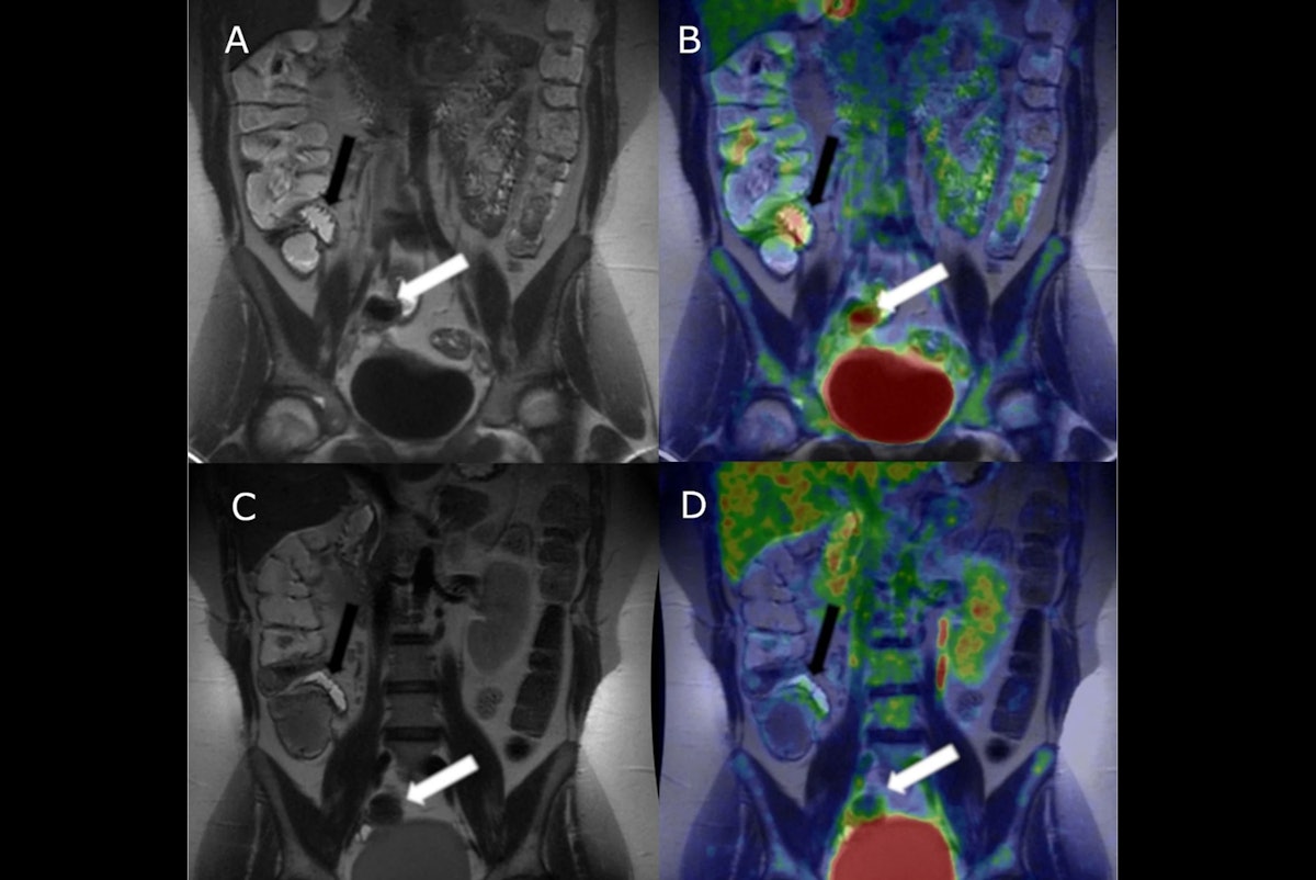
F-18 FDG-PET/MR enterography may be a useful tool for assessing responses to treatment in newly diagnosed patients with small bowel Crohn’s disease, researchers in Finland have reported.
The finding is from a study in which newly diagnosed patients underwent PET/MRE scans at baseline and then three months after starting standard treatments, noted lead author Juho Mattila, MD, a gastroenterologist at Turku University Hospital, and colleagues.
“These findings further support the potential of F-18 FDG-PET-MRE as a tool for evaluating intestinal inflammation in [Crohn’s disease],” the group wrote. The study was published August 26 in the European Journal of Nuclear Medicine and Molecular Imaging.
Crohn’s disease is a debilitating chronic inflammatory disease of the gastrointestinal tract; its global incidence is continuously rising, the authors noted. Both the diagnostics and follow-up of small bowel Crohn’s disease can be challenging, since it can be only partially evaluated by conventional endoscopy, they explained. While combined PET-MRE has shown potential in diagnosing small bowel disease, its role in monitoring treatment response has not been previously established, they added.
To that end, the group recruited 35 patients with suspected Crohn’s disease due to obscure diarrhea, abdominal pain, and elevated fecal calprotectin levels, or because they had a thickened bowel wall or stricture identified on abdominal CT. Prior to imaging, patients received injections of both F-18 FDG PET radiotracer and MRE gadolinium contrast agent. The scans took approximately 1.5 hours.
Participants were then referred to gastroenterologists, where 26 were diagnosed with small bowel Crohn’s disease and started on standard treatments such as corticosteroids or immune system suppressants. After three months, follow-up F-18 FDG-PET/MRE scans were performed in 18 (69%) of the patients.
According to the findings, median maximum standardized uptake values (SUVmax) of F-18 FDG radiotracer decreased significantly from baseline to follow-up (3.2 vs. 2.1, p = 0.0025). Meanwhile, the Simplified Magnetic Resonance Index of Activity (sMARIA) — an MRI-based scoring system used to measure the severity of the disease — was also significantly lower at follow-up (p = 0.001).
Notably, a reduction in SUVmax was observed in slightly more patients than a reduction in sMARIA (72% vs. 61%), which may suggest that resolution of inflammation is detectable earlier on PET imaging, the group added.
Ultimately, Crohn’s disease often leads to complications such as strictures, fistula, and abscesses, and diagnosing the disease and starting treatment early is essential, the researchers wrote. By combining quantitative information on glucose metabolism from PET sequences with detailed anatomical imaging from MRE sequences, clinicians can precisely assess the extent of inflammation, they suggested.
“Based on these results, F-18 FDG-PET/MRE appears to be a valuable tool not only for the diagnosis of small bowel [Crohn’s disease] but also for monitoring treatment response,” the group concluded.
The full study is available here.
