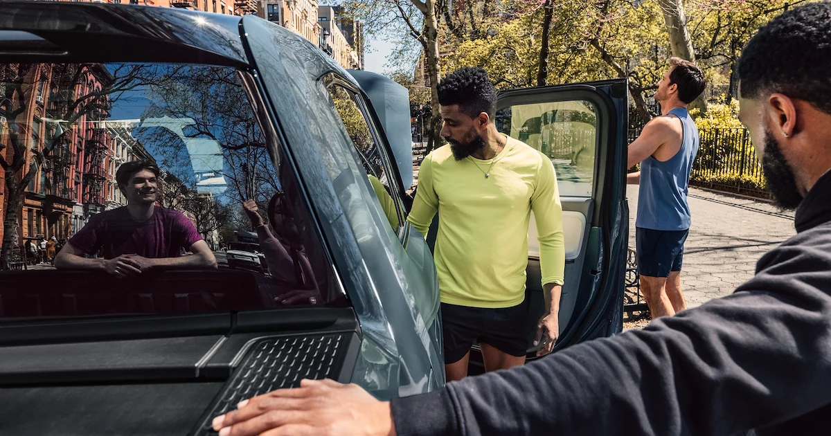
Introduction
Sodium-glucose cotransporter-2 (SGLT2) inhibitors are a relatively recent drug class that has been integrated into the therapeutic approach of Type 2 Diabetes Mellitus (Τ2DM) for over a decade. These agents exert their antihyperglycemic effect by acting on renal proximal convoluted tubules and inhibiting the SGLT2-mediated reabsorption of glucose; thus, they promote glucosuria irrespectively of plasma glucose concentration, which results in a daily urinary glucose excretion of 70–90 g when the therapeutic dosage is administered.1,2 Furthermore, SGLT2 inhibitors lower body weight and blood pressure, while having a beneficial impact on albuminuria and uric acid homeostasis.1,3,4 Large clinical trials have shown that therapy with SGLT2 inhibitors is associated with a lower risk of hospitalization for heart failure (HF), progression of renal disease, as well as all-cause and cardiovascular (CV) mortality.5–8 Recently, the cardiorenal protective effects of SGLT2 inhibitors have been expanded beyond diabetes, including patients with HF, regardless of ejection fraction,9–13 and people with chronic kidney disease (CKD), irrespective of the underlying cause.14–17 The fact that the cardiorenal benefit of SGLT-2 inhibitors emerges very early during the course of treatment and is observed independently of glycemic control and baseline glycated hemoglobin A1c (HbA1c) values indicates that these drugs act through pleiotropic mechanisms. The mechanisms responsible for these benefits remain incompletely understood. Several hypotheses have been proposed, including the “thrifty substrate hypothesis”, which suggests that SGLT2 inhibition promotes a shift toward more energy-efficient fuels such as ketone bodies, thereby improving mitochondrial efficiency and reducing oxidative stress. Such fuel energetics improvements may partially explain the observed clinical outcomes.
Metabolomics is a novel scientific field, developed to characterize comprehensively metabolites in a biological system, including plasma, serum, urine, tissue extracts, and cell cultures, in a high-throughput manner. The metabolome is defined as the amount of small endogenous molecules (molecular weight <1500 Da) that act as intermediates or end products of cellular metabolism, including lipids, organic acids, carbohydrates, amino acids, nucleotides, and steroids, representing a dynamic real-time view of the function of the cell or organism. Metabolomic analyses can be broadly divided into targeted and untargeted approaches. Targeted metabolomics measures predefined metabolites of interest with high quantitative accuracy and sensitivity, often focusing on specific pathways (eg, amino acids, lipids, ketones). In contrast, untargeted metabolomics aims to capture as many metabolites as possible in a biological sample, providing a global and unbiased overview of metabolic alterations, but often with lower sensitivity for individual metabolites and greater challenges in annotation and quantification.
The metabolic process is modulated by exogenous factors such as diet, lifestyle, or medication, and by endogenous inputs, such as age, sex, and gut microbiota-derived metabolites.18,19 Combining the identification of the metabolic phenotype of a biological matrix with advanced bioinformatics and analytical strategies, can be utilized to explore the pathophysiological pathways of complex disorders such as T2DM and CKD, discover new diagnostic or prognostic biomarkers, and investigate the biological influence of specific therapeutic interventions on disease progression.18,19 Key metabolite alterations, which have come across through metabolomics research regarding the metabolic footprint of T2DM and presented with consistency in the literature, include elevated plasma levels of branched-chain amino acids (BCAAs), aromatic amino acids, alpha-hydroxybutyrate and 2-aminoadipic acid. These changes may be evident before the onset of Τ2DM, in the prediabetic state, or in an overt disease.18,20–22 Conversely, reduced levels of glycine and lysophosphatidylcholine C18:2 have been noted both in individuals at high risk of T2DM and in patients with established disease.18
Experimental metabolomic studies in patients treated with SGLT2 inhibitors have demonstrated a metabolic shift away from glucose utilization toward alternative substrates, such as ketone bodies or amino acids, improvement in mitochondrial function, and changes in urine metabolite concentrations.23–26 However, existing studies are characterized by methodological heterogeneity and are often limited by small sample sizes and short follow-up periods. The lack of a comprehensive comparison between the metabolomic effects of each specific SGLT2 inhibitor hinders a complete understanding of their systemic effects and their broader metabolic implications.
The aim of this narrative review is to summarize and evaluate the existing metabolomic studies on different SGLT2 inhibitors in the context of T2DM, seeking to identify common pathways, class effects, and potential biomarkers that may contribute to the cardiorenal benefits of these agents.
Materials and Methods
The PubMed, Embase, and Google Scholar databases were searched to identify studies relevant to the metabolomic effect of SGLT2 inhibition in T2DM, regardless of publication status or year. The search strategy included combinations of the following keywords: “type 2 diabetes mellitus” OR “T2DM” OR “type 2 diabetes” AND “SGLT2” OR “SGLT2 inhibitors” OR “sodium-glucose cotransporter-2 inhibitors” OR “dapagliflozin” OR “empagliflozin” OR “canagliflozin” OR “ertugliflozin” OR “sotagliflozin” AND “metabolomics” OR “metabolome” OR “metabolic profiling” OR “metabolic signature”. The reference lists of all eligible studies and reviews were manually scanned for additional articles. Original studies in humans that investigated the metabolomic changes associated with the administration of SGLT2 inhibitors in patients with T2DM were selected for the review. Studies in animal models or in vitro settings were considered for background context, but were not included in the main synthesis. No language restrictions were applied during the initial search, but only articles published in English were examined. Additionally, we searched for studies involving ertugliflozin and the dual SGLT1/2 inhibitor sotagliflozin; however, eligible human metabolomics studies in T2DM were scarce to non-existent within our predefined criteria. Therefore, our synthesis focuses on dapagliflozin, empagliflozin, and canagliflozin.
Relevant articles were selected based on titles and abstracts, and full texts were reviewed to assess eligibility. The key findings of each included study were extracted and classified by each specific SGLT2 inhibitor.
Results
Metabolomic Effects of Dapagliflozin
Dapagliflozin is the compound in this class of drugs that has been studied the most, with several metabolomic investigations that underscoring its systemic metabolic impact beyond glycemic control. These studies have employed both targeted and untargeted metabolomic approaches in human subjects with T2DM, analyzing various biological matrices, including plasma, serum, and urine.
An untargeted mass spectrometry metabolomic analysis conducted by Mulder et al in 19 patients with T2DM and non-alcoholic fatty liver disease treated with dapagliflozin 10 mg/day with a 12-week follow-up period demonstrated significant changes in 108 plasma metabolites.27 More than 80% of the significantly increased metabolites belonged to the amino acid superpathway, while the remaining metabolites belonged to the xenobiotic, carbohydrates, and vitamins superpathways. N-acetyl aspartate (NAA), which is related to neuronal mitochondrial activity, was the amino acid in which the most prominent alterations were observed. Increases in BCAAs-derived carnitines, as well as metabolites related to histidine metabolism, the urea cycle, creatine, sarcosine and heme were also observed. On the other hand, most of the down-regulated metabolites were lipids, including diacylglycerols, endocannabinoids, dicarboxylic acids, and monohydroxylated fatty acids. Bile acids, plasma glucose and other simple carbohydrates, intermediates of urate, xanthine metabolism, and two amino acids, alanine and glutamine, were reduced after treatment, as well as intermediates in the tricarboxylic acid cycle (TCA), including succinate, fumarate, and malate. Significantly changed serum metabolites were further integrated with transcriptomic features measured in kidney tissues and analyzed for enriched pathways, revealing upregulation of citrulline metabolism superpathway, TCA cycle II, and L-carnitine biosynthesis, and decrease in glycine degradation (creatine biosynthesis). Dapagliflozin-induced changes, such as the increase in ketone bodies, short-chain acylcarnitines, and osmolytes (eg, betaine, myo-inositol), may contribute to improved renal energetics and cellular resilience. Elevated ketones and acylcarnitines provide more efficient mitochondrial substrates, potentially reducing renal hypoxia and oxidative stress. Meanwhile, osmolytes like betaine and myo-inositol help preserve tubular cell integrity under glucotoxic conditions. Collectively, these adaptations may underpin the observed renoprotective effects of dapagliflozin in patients with T2DM.
In another study by Mulder et al, the effects of dapagliflozin were investigated on a pre-specified panel of 13 urinary metabolites linked to mitochondrial function in 31 people with T2DM and albuminuria, using electron impact mass spectrometry.28 Regarding urine samples, 9 of 13 urinary metabolites were increased after 6 weeks of dapagliflozin treatment, resulting in a significant increase in the urinary metabolite index by 56% compared to placebo, while a significant increase in urine 3-hydroxybutyrate and urine acetoacetic acid was also observed.23 Since metabolites were found to be altered in urine but not plasma, it is suggested that these metabolites may reflect specific responses of renal tissue to SGLT2 inhibition. Considering the density of mitochondria in the kidneys, it has been hypothesized that amelioration of the mitochondrial function through reduction of excess glucose reabsorption and cellular workload, and selection of alternative fuel substrates, such as ketone bodies, improves tissue oxygenation, thus contributing to the renoprotective impact of dapagliflozin.
Proton nuclear magnetic resonance (1H-NMR) based metabolomics was used for another metabolomics study to evaluate the effect of dapagliflozin on serum metabolome in 50 patients with T2DM after 3 months of treatment.29 Regarding untargeted analysis, a good separation of metabolites was observed in the dapagliflozin group with a degree of overlap before and after treatment. In the targeted analysis, SGLT2 inhibition was shown to cause significant alterations in 10 of 46 identified metabolites; serum ketone bodies (3-hydroxy-butyrate, acetoacetate, acetone), tryptophane, citrate, and creatine increased, while serum glucose, taurine, threonine, and mannose levels decreased significantly after dapagliflozin administration. The authors postulated that ketones play a pivotal role in the beneficial cardiorenal profile of SGLT2 inhibitors, as they are considered a more efficient substrate for myocardial tissue compared to free fatty acids (FFAs) and glucose.30 Furthermore, they noted that an increase in citrate indicates an improvement in adenosine triphosphate (ATP) formation and thus in energy production,31 alteration of serum tryptophane levels may be associated with reduced inflammatory activity, and a decrease in mannose shows increased insulin sensitivity and improved insulin resistance.32,33
Bletsa et al evaluated the urine metabolic signature before and 3 months after the administration of dapagliflozin 10 mg/day in 50 patients with T2DM on monotherapy with metformin using 1H-NMR spectroscopy.26 Multivariate data analysis showed that the groups before and after dapagliflozin use were well separated with a small degree of overlap. Targeted analysis that followed demonstrated that 27 of the 70 quantified metabolites were significantly changed after SGLT2 inhibition; 2-aminobutyrate, 2-hydroxy-3-methylvalerate, 2-hydroxybutyrate, 2-hydroxyisovalerate, 3-hydroxybutyrate, 3-hydroxyisovalerate, acetoacetate, alanine, betaine, citrate, creatine, ethylmalonate, gluconate, glucose, hippurate, lactate, leucine, myo-inositol, N,N-dimethylglycine, N-methylhydantoin, sarcosine, trigonelline and valine were significantly increased, while chlorotyrosine, anserine, methanol and N-isovaleroylglycine were significantly reduced. Based on the results, the authors proposed that increased renal excretion of BCAAs is attributed to inhibition of SGLT2 and Na+/H+ exchanger (NHE3) transporters by dapagliflozin. Since amino acids participate in proximal tubular cell hypertrophy and, consequently, in diabetic nephropathy, reducing their proximal reabsorption may alleviate disease progression.34 Improvement in BCAA metabolism was further confirmed by the increase of six BCAA catabolism intermediates in urine samples from patients after dapagliflozin administration. A key finding of this study is the increase in betaine and myo-inositol in urine concentration after treatment. Beyond their roles as osmolytes, betaine and myo-inositol can contribute to the renoprotective effects of dapagliflozin. In previous studies, myo-inositol has been shown to protect renal cells from glucotoxicity and fibrosis,35 while betaine exerts anti-inflammatory properties36 and improves liver mitochondrial function.37 In addition, it was reported that the increase in the excretion of N-methylhydantoin, creatine, betaine, sarcosine, and N,N-dimethylglycin after therapy with dapagliflozin is associated with a potentially beneficial effect on gut microbiota metabolism.38 The significant increase induced by dapagliflozin in urine ketone bodies (3-hydroxybutyrate and acetoacetate) and lactate is also highlighted, as well as the alteration of citrate excretion, suggesting restored mitochondrial function and improved tubular energy metabolism.39
Liu et al conducted a multi-omics study, to explore the effect of dapagliflozin on the serum proteome and metabolome of 57 newly diagnosed patients with T2DM, who received 10mg/day of the agent for 12 weeks.40 In addition to a clear separation of metabolites before and after dapagliflozin administration in multivariate analysis, it was shown that SGLT2 inhibition significantly increased several amino acids, including aspartyl-leucine (Asp-Leu), aspartyl-isoleucine (Asp-Ile), aspartyl-phenylalanine (Asp-Phe), taurine and citrulline, and decreased 1,5-anhydroglucitol (1,5-AG), gluconolactone, ribose, hexose, succinic acid, xanthine and uric acid levels. The most differentially abundant metabolite was 1,5-AG levels – a polyol competing with glucose for renal reabsorption41 – indicating increased urinary glucose loss. The levels of other monosaccharides (ribose, hexose) and succinic acid [a TCA cycle intermediate42] also decreased, demonstrating altered glucose and energy metabolism. The negative energy balance induced by dapagliflozin was reflected in the increase in amino acids (Asp-Leu, Asp-Ile, and Asp-Phe), potentially indicating enhanced protein catabolism. Furthermore, taurine levels increased after treatment, a beneficial finding given the known functions of taurine in thermogenesis, mitochondrial function, and cardiac contractility.43 Consistent with previous studies,44,45 dapagliflozin reduced uric acid levels, possibly through enhanced urinary excretion, inhibition of xanthine oxidase, and suppression of the inflammatory response.46 The researchers suggest that reduced circulating levels of uric acid along with increased taurine levels may contribute to cardiorenal protection of dapagliflozin, while they emphasize that the significantly changed pathways of the combined analysis of proteins and metabolites are glycolysis/gluconeogenesis and the pentose phosphate pathway.
Two randomized placebo-control trials evaluated the association between dapagliflozin treatment and alterations in systemic metabolic pathways across the spectrum of HF using targeted mass spectrometry metabolite profiling in plasma samples; DEFINE-HF (Dapagliflozin Effects on Biomarkers, Symptoms and Functional Status in Patients With HF With Reduced Ejection Fraction)47 and PRESERVED-HF (Dapagliflozin in Preserved Ejection Fraction Failure).48 These studies did not recruit exclusively patients with T2DM; 64.5% (78 out of 121) and 54.7% (81 out of 148) of subjects in the dapagliflozin arm in DEFINE-HF and PRESERVED-HF, respectively, had T2DM. Selvaraj et al conducted a pooled analysis of the two studies that included 527 participants (59% with T2DM in the dapagliflozin group – 159 out of 269) and found that dapagliflozin administration significantly affected metabolite clusters enriched in ketone-related metabolites and short-/medium-chain acylcarnitines, compared to placebo.49 While short-/medium-chain acylcarnitines increased consistently in all values of left ventricular ejection fraction (LVEF), ketone metabolism biomarkers decreased at higher LVEF. Amino acids, including BCAAs, did not show any significant trend. Since the substrate shift towards ketone utilization is a cornerstone of HF pathophysiology, especially in HF with reduced ejection fraction (HFrEF), the mild raise in ketone levels induced by dapagliflozin may imply the role of SGLT2 inhibition in metabolic reprogramming of the disease and thus, explaining in part the observed CV benefit.50,51 It was also hypothesized that the increase in short- and medium-chain acylcarnitines—without changes in long-chain ones—may indicate improved mitochondrial function and enhanced fatty acid oxidation, which has been associated with improved myocardial energetics and contractility.52
Interestingly, another study explored the metabolic effects of SGLT2 inhibitors in diabetes-associated HF with preserved ejection fraction (HFpEF) in 20 individuals with T2DM and HFpEF treated with dapagliflozin for more than 3 months.53 Through serum metabolome analysis, long-term administration of dapagliflozin showed significant changes in metabolites related to nicotinamide metabolism, arginine biosynthesis, and cyclic adenosine monophosphate (cAMP) and estrogen signaling pathways. Notably, nicotinamide, a key component of the coenzymes nicotinamide adenine dinucleotide (NAD) and nicotinamide adenine dinucleotide phosphate (NADP), plays a critical role in mitochondrial respiratory chain oxidation and energy production, suggesting a dapagliflozin-induced improvement in mitochondrial function and energetics of cardiomyocytes. These findings offer molecular insights into how SGLT2 inhibitors may refine myocardial injury, independently of their antihyperglycemic action.
Metabolomic Effects of Empagliflozin
The metabolic signature of empagliflozin was investigated in a prospective study by Kappel et al, including 25 patients with T2DM and established CV disease, who were treated with empagliflozin 10 mg/day for one month.24 Using untargeted serum metabolomics and specifically liquid chromatography‒tandem mass spectrometry, it was found that SGLT2 inhibition significantly affected 162 metabolites. Empagliflozin treatment led to reductions in glucose and other carbohydrates, while increased levels of aconitate and fumarate indicated activation of the TCA cycle. Furthermore, an increase in short-chain but not long- or very long-chain acylcarnitines suggested enhanced ATP production through degradation of fatty acids, amino acids, and ketone bodies, rather than through triglyceride breakdown. Pathway enrichment analysis further revealed that increased catabolism of ketogenic amino acids and BCAAs (lysine, leucine, isoleucine, and valine) induced by empagliflozin, increased TCA cycle, and ketogenesis. Another key finding of this study was the elevation of urea cycle intermediates, implying increased amino acid catabolism and nitrogen handling. Since the utilization of ketone bodies plays a critical role in cardiomyocyte energy production and further in mitochondrial function in HF in the context of diabetes due to impaired glucose and fatty acid oxidation, the authors underscore that the observed fuel selection towards ketones and BCAAs provoked by empagliflozin may contribute to the cardioprotective effects of this agent.30,54
The potential association between the proposed efficiency of ketone bodies as a substrate for the failing heart and the mild hyperketonemia observed with SLGT2 inhibitors was further evaluated as an exploratory endpoint in the EMPA-VISION trial (Assessment of Cardiac Energy Metabolism, Function and Physiology in Patients trial With Heart Failure Taking Empagliflozin).55,56 In this randomized, placebo-controlled study, 72 patients with HFrEF (n = 36) or HFpEF (n = 36) were enrolled and randomly assigned to receive 10 mg empagliflozin or placebo once daily for 12 weeks. Only 12.55% of the total study population had concomitant T2DM. Mass spectrometry and principal component analysis were used to assess the impact of empagliflozin administration on a set of 19 targeted metabolites related to energy metabolism.57 However, no significant differences in serum metabolites between drug and placebo were identified. Circulating levels of ketone bodies (β-hydroxybutyrate) and FFAs remained unchanged after empagliflozin treatment. Moreover, the study failed to detect measurable improvements in cardiac energetics after SGLT2 inhibition in patients of either group, suggesting that mechanisms beyond the thrifty fuel hypothesis may account for the beneficial effects of empagliflozin observed across the spectrum of HF.30,58,59 As previously mentioned, the so-called “thrifty fuel hypothesis” suggests that a shift toward more energy-efficient substrates such as ketone bodies may enhance cardiac and renal energetics. Clinical and experimental studies indicate that ketones produce more ATP per unit of oxygen consumed compared to glucose or fatty acids, thereby improving mitochondrial efficiency and reducing oxidative stress. This metabolic adaptation may contribute to the observed cardio-renal benefits of SGLT2 inhibitors, supporting the clinical relevance of the thrifty fuel hypothesis.
Approaching metabolomics as a tool for biomarker identification, Kappel et al combined three different untargeted serum metabolomics datasets in both human and mouse models to discover metabolites related to a diabetic phenotype in two prospective cohorts; 37 patients with and without T2DM hospitalized for myocardial infarction and 25 patients with T2DM included in an empagliflozin registry.60 At baseline, 1,5-AG levels were inversely correlated with HbA1c and serum glucose, effectively distinguishing between varying degrees of glycemic control. However, following empagliflozin treatment, 1,5-AG levels decreased significantly in all patient groups, regardless of HbA1c levels, thus diminishing its correlation with traditional glycemic markers. This effect was also observed in mice with diabetes treated with empagliflozin. Subsequently, the study group conducted a targeted serum analysis of 1,5-AG in the setting of a placebo‑controlled, randomized, double-blind human trial with empagliflozin at baseline, 72 hours and 3 months after treatment (n=42), which confirmed empagliflozin-induced reduction in 1,5-AG levels, possibly due to increased urinary excretion. Therefore, while 1,5-AG is a useful indicator of glycemic control, a limited utility is proposed in patients on treatment with SGLT2 inhibitors.41
A novel study explored the hypothesis that the CV benefits of empagliflozin could be associated with alterations in the gut microbiota and plasma metabolites. In this trial, 76 treatment naive patients with T2DM and risk factors for CV disease were randomized to receive empagliflozin 10 mg/day or metformin 1700 mg/day for 3 months.61 Untargeted serum metabolomics analysis revealed a change in 27 metabolites after empagliflozin administration; the most prominent was the significant increase in sphingomyelin and capric acid levels and the decrease in glycochenodeoxycholate, cis-aconitate, erythritol and uric acid. According to evidence, sphingomyelin can prevent the translocation of gut bacteria-derived lipopolysaccharide and inhibit its pro-inflammatory effects.62 In addition to metabolomics, it should be highlighted that empagliflozin increased beneficial short-chain fatty acid (SCFA) producing bacteria, including Roseburia, Eubacterium, and Faecalibacterium, while reducing potentially harmful species such as Escherichia-Shigella, Bilophila, and Hungatella. Therefore, it is speculated that SGLT2 inhibition may lead to an improved intestinal microbiome profile and that this pathway may represent another mechanism of action of these agents.63
Metabolomic Effects of Canagliflozin
Canagliflozin is a less selective SGLT2 inhibitor, which can also partially inhibit SGLT1, reducing this way not only renal, but also intestinal glucose absorption.64 Canagliflozin has demonstrated significant renoprotective and cardiometabolic effects in patients with T2DM.1,7 Compared to dapagliflozin or empagliflozin, fewer metabolomic studies have focused on canagliflozin, while the majority have been performed in animal models or cell cultures. However, emerging evidence highlights distinct metabolic adaptations associated with canagliflozin administration, particularly in the context of CKD.
Shao et al performed metabolomic analyses in plasma samples from 31 patients with T2DM and CKD, treated with canagliflozin 100 mg/day for 12 weeks.65 The untargeted assay revealed suppression of glycolysis and upregulation of fatty acid oxidation, while upregulation of glycine was observed by targeted metabolomics. Further in vitro experiments demonstrated that glycine levels increased and associated pathways, such as glycine, serine, and threonine metabolism, were significantly up-regulated in canagliflozin-treated kidneys. Glycine supplementation in db/db mice and human proximal tubular epithelial cells improved insulin sensitivity, decreased blood glucose, and alleviated renal damage, likely through activation of the AMP-activated protein kinase (AMPK)/mammalian target of the rapamycin (mTOR) signaling pathway. Metabolomic profiling exhibited a metabolic shift resembling fasting- and aestivation-like states, which can relieve mitochondrial overload and oxidative stress. These findings suggest that the renoprotective effects of canagliflozin can be attributed to metabolic reprogramming that enhances energy efficiency and reduces cellular stress in the kidneys.
Taken together, these studies reveal both shared and distinct metabolomic signatures across SGLT2 inhibitors. Dapagliflozin is most consistently associated with shifts toward ketone metabolism and enhanced amino acid catabolism. Empagliflozin shows more variable effects, particularly with regard to cardiac energetics, reflecting differences in study design and populations. Canagliflozin has fewer studies but appears to share the class-wide features of enhanced ketogenesis and modulation of amino acid metabolism. This comparative synthesis highlights both class effects and compound-specific nuances in metabolic adaptation. We did not identify eligible human metabolomics studies in T2DM for ertugliflozin or sotagliflozin; thus, results are summarized for dapagliflozin, empagliflozin, and canagliflozin. A summary of the main characteristics and metabolomic findings of the human studies reviewed is provided in Table 1.
Table 1 Summary of Human Metabolomics Studies Investigating SGLT2 Inhibitors
|
Interpretation of findings must take into account methodological differences across studies. Variations in sample type (serum, plasma, urine), analytical platforms (NMR, ultra-performance liquid chromatography coupled with mass spectrometry, gas chromatography coupled with mass spectrometry), and study populations (healthy vs T2DM, variable baseline renal function) likely contribute to heterogeneity in results. In some cases, targeted approaches captured small but clinically meaningful changes (eg, 1,5-AG), while untargeted studies revealed broader pathway-level shifts. These methodological distinctions may explain discrepancies in metabolite reporting across studies.
Discussion
To our knowledge, this is the first narrative review in the literature to summarize current evidence from human studies on the metabolic effects of SGLT2 inhibitors in T2DM. These drugs, originally developed for their glucose-lowering capacity, have consistently demonstrated cardiorenal protective actions in populations with or without diabetes. Metabolomics is a novel approach to explore metabolic changes related to disease development and progression, and has emerged as a valuable tool to discover the molecular pathways through which SGLT2 inhibitors exert their pleiotropic benefits. Although comparing results from independent metabolomic studies is challenging, this research field has offered insights into key metabolic adaptations associated with SGLT2 inhibition therapy. While each compound presents with unique features and specific pharmacological properties, several overlapping mechanisms may account for their protective effects on the heart and kidneys, which have been identified as class effects in large randomized trials.
Common metabolic signatures observed across the agents examined, (dapagliflozin, empagliflozin, and canagliflozin), include the induction of a metabolic shift away from glucose utilization toward alternative and more energy-efficient fuel sources, such as ketone bodies, fatty acids, and certain amino acids. In most studies, SGLT2 inhibitors have been shown to promote ketone body production, upregulate fatty acid oxidation, and facilitate the breakdown of branched chain and ketogenic amino acids, along with reductions in glucose, uric acid, and certain amino acids such as alanine and glutamine.
SGLT2 inhibitors lead to glycosuria and reduction of plasma glucose levels, which in turns results in decrease in insulin-to-glucagon ration, augmented lipolysis and a mild increase in hepatic production of ketone bodies.1 Among the most consistently observed and biologically relevant metabolic changes, following treatment with SGLT2 inhibitors, was the rise in circulating ketone bodies. While FFAs are the primary fuel for the healthy myocardium, carbohydrates, although energy-inefficient, become the main energy source for the failing heart.30 Since ketone bodies oxidation produces more energy than glucose oxidation, it has been proposed that hyperketonemia and increase in FFAs induced by SGLT2 inhibitors drive the myocardial tissue to selectively use ketone bodies and FFAs; this shift to more potent energy substrates improves energy deficit and further cardiac energetics and function, contributing to the risk reduction in HF outcomes seen with this class of drugs. In the pooled analysis of DEFINE-HF and PRESERVED-HF,49 dapagliflozin was associated with elevated ketone bodies and short-/medium-chain acylcarnitines, which was in line with findings in studies using HFrEF animal models.66,67 The robustness of the study by Selvaraj et al is supported by its large sample size, inclusion of a placebo arm, and identification of a wide range of metabolites, as well as inclusion of data from two trials on the effects of dapagliflozin in people with HF.49 Similarly, empagliflozin administration was able to enhance ketogenesis and increase short-chain acylcarnitines in the study by Kappel et al, who evaluated this effect in patients with T2DM and CV disease.24 However, the EMPA-VISION trial did not detect significant alterations in circulating levels of ketone bodies and FFAs after treatment with empagliflozin.55 The authors attributed this difference mainly to the composition of the study populations; EMPA-VISION enrolled only patients with non-ischemic HFrEF, while more than half of DEFINE-HF subjects presented with ischemic HFrEF. Ischemia and subsequently, decreased oxygen supply, may modify metabolic profile and fuel utilization, even before the onset of HF.68 It has to be emphasized that only 12.5% of patients in the EMPA-VISION trial had T2DM, which may have limited the ketone response observed, as individuals with diabetes typically exhibit a more pronounced shift toward ketogenesis under SGLT2 inhibition. Although the degree of elevation of ketones varied between studies and was not universally detected, the consistent trend suggests a potential mechanistic association between SGLT2 inhibition and improved cardiac outcomes by metabolic reprogramming.
The elevation of ketone bodies observed with SGLT2 inhibition may provide a more efficient myocardial and renal fuel source, supporting improved energetics in conditions such as HF and CKD. Clinical data showing associations between higher ketone availability and improvements in ejection fraction and reduced albuminuria lend correlative support to this mechanism. However, not all studies align with the “thrifty substrate hypothesis”; for instance, Hundertmark et al55 reported no clear improvement in cardiac energetics in patients with T2DM treated with empagliflozin, underscoring that the relationship between metabolite shifts and clinical endpoints remains partially speculative and may differ by compound and patient population.
SGLT2 inhibitors appear to promote broader metabolic alterations, involving changes in amino acid metabolism and mitochondrial function. Increased TCA cycle activity suggests improved mitochondrial function and oxidative phosphorylation efficiency, ameliorating this way oxidative stress at the cellular level and enhancing energy production. Of interest, decreased levels of TCA metabolites were observed by Mulder et al after dapagliflozin treatment,27 which is in contrast to the empagliflozin-induced effect in the study by Kappel et al.24 The authors attributed this finding to possible differences in the study population or duration of treatment between the two trials, as well as to increased gluconeogenesis and improved mitochondrial efficacy, reflected by a greater degree of complete fatty acid oxidation. SGLT2 inhibition also restored the accumulation of TCA intermediates in kidneys in another study that used animal models.69 As already mentioned, low plasma glycine levels have been associated with diabetes and obesity.18 Canagliflozin was shown to upregulate glycine, serine, and threonine pathways in patients with T2DM and CKD,65 which was also demonstrated in another study with db/db mice treated with empagliflozin.70 Considering the osmotic diuresis and water loss caused by SGLT2 inhibition, it has been hypothesized that an aestivation-like mechanism underpins the renal protective effects of these agents.71 Estivators produce high levels of osmolytes in order to compensate for energy and water loss, and amino acids offer the necessary energy and nitrogen for this process. The observed upregulation of amino acid catabolism appears to be a component of the broader metabolic reprogramming induced by SGLT2 inhibition, rather than its primary cause. This reprogramming reflects a coordinated shift in substrate utilization toward amino acids, ketones, and fatty acids as alternative energy sources. In addition, the administration of dapagliflozin and empagliflozin was associated with increased BCAA breakdown and improved protein catabolism. The resulting metabolic adaptations indicate that SGLT2 inhibition may alleviate cellular stress by promoting substrate flexibility and reducing mitochondrial overload, particularly in insulin-resistant and hypoxic tissues such as the diabetic kidney and failing myocardium.
Another intriguing finding related to amino acid metabolism is the inconsistency reported in taurine levels following dapagliflozin use. While Liu et al found increased serum taurine concentrations following treatment,40 another study observed that dapagliflozin significantly decreased taurine levels.29 The authors of the latter study suggested that the discrepancy may be explained, at least in part, by the absence of a control arm in the study by Liu et al, which limits the interpretation of treatment effects. In addition, they proposed that the decrease in serum taurine may result from enhanced urinary excretion triggered by SGLT2 inhibition.
Recent findings highlight the potential role of the gut microbiota in mediating some of the beneficial effects of SGLT2 inhibitors. Alterations in intestinal microbial composition and metabolite production have been observed following treatment with empagliflozin.61 On the one hand, elevated levels of SCFA-producing bacteria were detected, which have been shown to regulate blood glucose levels, reduce oxidative stress and exert anti-inflammatory and antitumorigenic activity.72 According to current evidence, SCFAs improved CV-related metabolic disorders in diabetic mice, and therefore, they have emerged as a promising strategy for CV disease prevention.73 On the other hand, empagliflozin administration decreased harmful species, related to inflammation, disruption of the gut barrier, insulin resistance, hyperglycemia, and adverse CV outcomes.74–76 In contrast to these results, another double-blind randomized trial, comparing dapagliflozin to gliclazide administration in patients with T2DM, failed to demonstrate significant alterations in fecal microbiota composition.77 The inconsistent results between these two trials can be attributed to differences in the selected interventions, as well as variations in patient characteristics and methodologies used for gut microbiome analysis. Interestingly, Bletsa et al reported that the increase in specific metabolites in urine, such as N-methylhydantoin, creatine, betaine, sarcosine, and N,N-dimethylglycin, in response to dapagliflozin treatment, may be associated with a beneficial effect on gut microbiota metabolism.26 These findings suggest that beyond glucose control, SGLT2 inhibitors may exert part of their pleiotropic actions through crosstalk between the gut and distant organs, particularly in the context of CV disease.
Chronic low-grade inflammation is a pathophysiological hallmark of T2DM and CV disease. In the study by Filippas-Ntekouan et al, it was shown that serum tryptophane levels increased in response to dapagliflozin treatment and this finding was associated with reduced inflammatory activity.29 The rationale behind this speculation is that tryptophane is converted to kynurenine by the rate-limiting enzyme indoleamine 2,3-dioxygenase (IDO), which is upregulated by proinflammatory molecules such as interferon‐gamma (IFN-γ) and interleukins. A reduced kynurenine/tryptophane ratio indicates reduced IDO activity and, by extension, reduced systemic inflammation. Therefore, the observed increase in tryptophan levels and indirect reduction in IDO activity after dapagliflozin administration can be attributed to a suppression of proinflammatory signaling induced by SGLT2 inhibition. These findings are in accordance with previous data proposing that SGLT2 inhibitors may exert part of their cardiometabolic benefits through anti-inflammatory mechanisms.78–80 Additionally, the anti-inflammatory properties of betaine, may further contribute to the renoprotective effects of dapagliflozin, as suggested by changes in its urinary excretion profile.26 From a translational perspective, several metabolites emerging from these studies could serve as biomarkers for SGLT2 inhibitor response. Reductions in 1,5-AG may reflect improved glycemic control, while elevations in acylcarnitines and BCAAs could provide early indicators of metabolic reprogramming. Such biomarkers hold potential for use in precision medicine, enabling the prediction or monitoring of therapeutic efficacy in individual patients.
Despite the valuable insights gained from current metabolomic studies, several limitations must be acknowledged. First of all, the majority of studies include a relatively small sample size, which limits statistical power and generalizability of the results. Short follow-up periods also hamper the ability to detect long-term metabolic changes. Second, there is substantial heterogeneity in the study design in terms of demographics, patient characteristics such as presence or absence of diabetes, preserved or impaired renal function and HF phenotype, differences in the type and dose of SGLT2 inhibitor, as well as duration of treatment, which hinders direct comparison and meta-analysis of the findings of various studies. Moreover, few studies have incorporated control arms and randomization; therefore, the observed alterations cannot be exclusively attributed to the intervention. What also makes direct comparisons between studies challenging is the discrepancy in analytical platforms and metabolite panels used. More specifically, while untargeted metabolomics can provide broader information, this is often limited by issues of metabolite identification, quantification accuracy, and biological interpretability. However, targeted validation cannot assay all metabolic pathways affected by SGLT2 inhibition. Another important consideration is that most of the findings are derived from animal models or in vitro experiments, and human evidence remains relatively limited. Although associations between metabolic changes and clinical outcomes are frequently observed, causality has not been established and remains speculative. Since metabolomic studies serve as a tool for identifying a multitude of metabolites, it is intuitively understood why the complete characterization of the underlying mechanisms and potential consequences is extremely difficult. Finally, most studies focus on serum or urine samples, whereas tissue-related metabolomic changes are underexplored – for example, urine metabolites may not necessarily be specific to kidney energetics. In either case, metabolomic analyses provide only snapshots of metabolic physiology and may not capture dynamic tissue-specific changes over time. However, our review summarizes the available evidence from human studies that elucidates the effect of SGLT2 inhibition on the metabolome in the context of T2DM and establishes a comprehensive reference point for future investigations.
Future research should employ large-scale longitudinal studies that will also incorporate randomized, controlled designs and head-to-head comparisons of different SGLT2 inhibitors to distinguish class effects from compound-specific actions. Reproducibility and accuracy could be achieved through standardized workflow protocols that include sample collection, data processing, and analysis, in parallel with the combination of untargeted and targeted metabolomic assays. Integrating metabolomics with other omics platforms, such as transcriptomics, proteomics, and microbiome sequencing, may lead to a more robust association between modulation of metabolic pathways and inhibition of SGLT2. Furthermore, future studies should aim to identify predictive biomarkers of treatment response and assess the potential correlation of metabolic changes with clinical endpoints, thus contributing to the implementation of molecular findings into clinical practice and further into precision medicine. Future work should address the current gap by evaluating ertugliflozin, sotagliflozin and other molecules using harmonized untargeted and targeted metabolomic approaches to delineate class-wide versus compound-specific metabolic signatures. Finally, expanding research beyond T2DM — into HF phenotypes, CKD, and populations without diabetes — will broaden the clinical impact of these insights.
Conclusion
Metabolomic profiling has identified a network of metabolic adaptations induced by SGLT2 inhibitors in T2DM, beyond their glucose-lowering action. These agents are suggested to exert potent cardiometabolic and renoprotective effects by promoting ketogenesis, enhancing fatty acid and amino acid catabolism, modulating mitochondrial function, and attenuating inflammation. Although current human studies provide valuable mechanistic perspectives, more research through standardized, multi-omic investigations and head-to-head comparisons will be essential to fully elucidate compound-specific pathways and identify biomarkers predictive of treatment response. Such advances will refine therapeutic strategies and pave the way to personalized medicine in diabetes and its complications.
Abbreviations
1,5-AG, 1,5-anhydroglucitol; AMPK, AMP-activated protein kinase; Asp-Ile, Aspartyl-Isoleucine; Asp-Leu, Aspartyl-Leucine; Asp-Phe, Aspartyl-Phenylalanine; ATP, Adenosine triphosphate; BCAA, Branched-chain amino acid; cAMP, Cyclic adenosine monophosphate; CKD, Chronic Kidney Disease; CV, Cardiovascular; FFAs, Free Fatty Acids; HbA1c, Glycated Hemoglobin A1c; 1H-NMR, Proton Nuclear Magnetic Resonance; HF, Heart Failure; HFpEF, Heart Failure with Preserved Ejection Fraction; HFrEF, Heart Failure with Reduced Ejection Fraction; IDO, Indoleamine 2,3-dioxygenase; IFN-γ, Interferon‐gamma; LVEF, Left Ventricular Ejection Fraction; mTOR, Mammalian target of rapamycin; NAA, N-acetyl aspartate; NAD, Nicotinamide Adenine Dinucleotide; NADP, Nicotinamide Adenine Dinucleotide Phosphate; NHE3, Na+/H+ exchanger transporter; SCFA, Short-chain fatty acid; SGLT2, Sodium-glucose co-transporters 2; T2DM, Type 2 Diabetes Mellitus; TCA, Tricarboxylic acid cycle.
Author Contributions
All authors made a significant contribution to the work reported, whether that is in the conception, study design, execution, acquisition of data, analysis and interpretation, or in all these areas; took part in drafting, revising or critically reviewing the article; gave final approval of the version to be published; have agreed on the journal to which the article has been submitted; and agree to be accountable for all aspects of the work.
Funding
No funding was received for the conduction of this study.
Disclosure
TK has received honoraria for lectures/advisory boards and research support from AstraZeneca, Boehringer Ingelheim, Pharmaserve Lilly, Sanofi, ELPEN, Menarini and Novo Nordisk. KK has received honoraria for lectures/advisory boards and research support from Astra Zeneca, Boehringer Ingelheim, Pharmaserve Lilly, Sanofi-Aventis, ELPEN, MSD and Novo Nordisk. Other authors report no competing interest in this work.
References
1. Brown E, Rajeev SP, Cuthbertson DJ, Wilding JPH. A review of the mechanism of action, metabolic profile and haemodynamic effects of sodium-glucose co-transporter-2 inhibitors. Diabetes Obes Metab. 2019;21(S2):9–18. doi:10.1111/dom.13650
2. Rajeev SP, Cuthbertson DJ, Wilding JPH. Energy balance and metabolic changes with sodium-glucose co-transporter 2 inhibition. Diabetes Obes Metab. 2016;18(2):125–134. doi:10.1111/DOM.12578
3. DeFronzo RA, Norton L, Abdul-Ghani M. Renal, metabolic and cardiovascular considerations of SGLT2 inhibition. Nat Rev Nephrol. 2017;13(1):11–26. doi:10.1038/NRNEPH.2016.170
4. Monami M, Nardini C, Mannucci E. Efficacy and safety of sodium glucose co-transport-2 inhibitors in type 2 diabetes: a meta-analysis of randomized clinical trials. Diabetes Obes Metab. 2014;16(5):457–466. doi:10.1111/DOM.12244
5. Zinman B, Wanner C, Lachin JM, et al. Empagliflozin, cardiovascular outcomes, and mortality in type 2 diabetes. N Engl J Med. 2015;373(22):2117–2128. doi:10.1056/NEJMoa1504720
6. Wiviott SD, Raz I, Bonaca MP, et al. Dapagliflozin and cardiovascular outcomes in type 2 diabetes. N Engl J Med. 2019;380(4):347–357. doi:10.1056/NEJMOA1812389
7. Neal B, Perkovic V, Mahaffey KW, et al. Canagliflozin and cardiovascular and renal events in type 2 diabetes. N Engl J Med. 2017;377(7):644–657. doi:10.1056/NEJMOA1611925
8. Zelniker TA, Wiviott SD, Raz I, et al. SGLT2 inhibitors for primary and secondary prevention of cardiovascular and renal outcomes in type 2 diabetes: a systematic review and meta-analysis of cardiovascular outcome trials. Lancet. 2019;393(10166):31–39. doi:10.1016/S0140-6736(18)32590-X
9. Solomon SD, McMurray JJV, Claggett B, et al. Dapagliflozin in heart failure with mildly reduced or preserved ejection fraction. N Engl J Med. 2022;387(12):1089–1098. doi:10.1056/NEJMOA2206286
10. McMurray JJV, Solomon SD, Inzucchi SE, et al. Dapagliflozin in patients with heart failure and reduced ejection fraction. N Engl J Med. 2019;381(21):1995–2008. doi:10.1056/NEJMOA1911303
11. Anker SD, Butler J, Filippatos G, et al. Empagliflozin in heart failure with a preserved ejection fraction. N Engl J Med. 2021;385(16):1451–1461. doi:10.1056/NEJMOA2107038
12. Packer M, Anker SD, Butler J, et al. Cardiovascular and renal outcomes with empagliflozin in heart failure. N Engl J Med. 2020;383(15):1413–1424. doi:10.1056/NEJMOA2022190
13. McDonagh TA, Metra M, Adamo M, et al. 2023 focused update of the 2021 ESC guidelines for the diagnosis and treatment of acute and chronic heart failure developed by the task force for the diagnosis and treatment of acute and chronic heart failure of the European Society of Cardiology (ESC) with the special contribution of the Heart Failure Association (HFA) of the ESC. Eur Heart J. 2023;44(37):3627–3639. doi:10.1093/EURHEARTJ/EHAD195
14. Stevens PE, Ahmed SB, Carrero JJ, et al. KDIGO 2024 clinical practice guideline for the evaluation and management of chronic kidney disease. Kidney Int. 2024;105(4):S117–S314. doi:10.1016/J.KINT.2023.10.018
15. Perkovic V, Jardine MJ, Neal B, et al. Canagliflozin and renal outcomes in type 2 diabetes and nephropathy. N Engl J Med. 2019;380(24):2295–2306. doi:10.1056/nejmoa1811744
16. Heerspink HJL, Stefánsson BV, Correa-Rotter R, et al. Dapagliflozin in patients with chronic kidney disease. N Engl J Med. 2020;383(15):1436–1446. doi:10.1056/nejmoa2024816
17. The EMPA-KIDNEY Collaborative Group. Empagliflozin in patients with chronic kidney disease. N Engl J Med. 2023;388(2):117–127. doi:10.1056/NEJMoa2204233
18. Klein MS, Shearer J. Metabolomics and type 2 diabetes: translating basic research into clinical application. J Diabetes Res. 2016;2016:3898502. doi:10.1155/2016/3898502
19. Sas KM, Karnovsky A, Michailidis G, Pennathur S. Metabolomics and diabetes: analytical and computational approaches. Diabetes. 2015;64(3):718–732. doi:10.2337/DB14-0509
20. Newgard CB. Metabolomics and metabolic diseases: where do we stand? Cell Metab. 2016;25(1):43. doi:10.1016/J.CMET.2016.09.018
21. Gall WE, Beebe K, Lawton KA, et al. alpha-hydroxybutyrate is an early biomarker of insulin resistance and glucose intolerance in a nondiabetic population. PLoS One. 2010;5(5). doi:10.1371/JOURNAL.PONE.0010883
22. Wang TJ, Ngo D, Psychogios N, et al. 2-Aminoadipic acid is a biomarker for diabetes risk. J Clin Invest. 2013;123(10):4309–4317. doi:10.1172/JCI64801
23. Mulder S, Heerspink HJL, Darshi M, et al. Effects of dapagliflozin on urinary metabolites in people with type 2 diabetes. Diabetes Obes Metab. 2019;21(11):2422–2428. doi:10.1111/dom.13823
24. Kappel BA, Lehrke M, Schütt K, et al. Effect of empagliflozin on the metabolic signature of patients with type 2 diabetes mellitus and cardiovascular disease. Circulation. 2017;136(10):969–972. doi:10.1161/CIRCULATIONAHA.117.029166
25. Jo HA, Seo JH, Lee S, et al. Metabolomic profiling in kidney cells treated with a sodium glucose-cotransporter 2 inhibitor. Sci Rep. 2023;13(1):1–8. doi:10.1038/s41598-023-28850-3
26. Bletsa E, Filippas-Dekouan S, Kostara C, et al. Effect of dapagliflozin on urine metabolome in patients with type 2 diabetes. J Clin Endocrinol Metab. 2021;106(5):1269–1283. doi:10.1210/CLINEM/DGAB086
27. Mulder S, Hammarstedt A, Nagaraj SB, et al. A metabolomics-based molecular pathway analysis of how the sodium-glucose co-transporter-2 inhibitor dapagliflozin may slow kidney function decline in patients with diabetes. Diabetes Obes Metab. 2020;22(7):1157–1166. doi:10.1111/dom.14018
28. Sharma K, Karl B, Mathew AV, et al. Metabolomics reveals signature of mitochondrial dysfunction in diabetic kidney disease. J Am Soc Nephrol. 2013;24(11):1901–1912. doi:10.1681/ASN.2013020126
29. Filippas-Ntekouan S, Dimou A, Dafopoulos P, et al. Effect of dapagliflozin on the serum metabolome in patients with type 2 diabetes mellitus. J Diabetes Metab Disord. 2024;24(1):4. doi:10.1007/s40200-024-01508-1
30. Ferrannini E, Mark M, Mayoux E. CV protection in the EMPA-REG OUTCOME trial: a thrifty substrate hypothesis. Diabetes Care. 2016;39(7):1108–1114. doi:10.2337/DC16-0330
31. Zhang X, Wang N, Fu P, et al. Dapagliflozin attenuates heart failure with preserved ejection fraction remodeling and dysfunction by elevating β-hydroxybutyrate-activated citrate synthase. J Cardiovasc Pharmacol. 2023;82(5):375–388. doi:10.1097/FJC.0000000000001474
32. Fortin E, Campi B, Ferrannini E, et al. High mannose correlates with surrogate indexes of insulin resistance and is associated with an increased risk of cardiovascular events independently of glycemic status and traditional risk factors. Diabetes Care. 2024;47(2):246–251. doi:10.2337/DC23-0870
33. Fakhrolmobasheri M, Abhari AP, Manshaee B, et al. Effect of sodium–glucose cotransporter 2 inhibitors on insulin resistance; a systematic review and meta-analysis. Acta Diabetol. 2023;60(2):191–202. doi:10.1007/S00592-022-01981-1
34. Vallon V, Gerasimova M, Rose MA, et al. SGLT2 inhibitor empagliflozin reduces renal growth and albuminuria in proportion to hyperglycemia and prevents glomerular hyperfiltration in diabetic Akita mice. Am J Physiol – Ren Physiol. 2014;306(2):194–204. doi:10.1152/AJPRENAL.00520.2013
35. Chang HH, Chao HN, Walker CS, Choong SY, Phillips A, Loomes KM. Renal depletion of myo-inositol is associated with its increased degradation in animal models of metabolic disease. Am J Physiol – Ren Physiol. 2015;309(9):F755–F763. doi:10.1152/AJPRENAL.00164.2015
36. Zhao G, He F, Wu C, et al. Betaine in inflammation: mechanistic aspects and applications. Front Immunol. 2018;9:370622. doi:10.3389/FIMMU.2018.01070
37. Zhang L, Qi Y, Aluo Z, Liu S, Zhang Z, Zhou L. Betaine increases mitochondrial content and improves hepatic lipid metabolism. Food Funct. 2019;10(1):216–223. doi:10.1039/C8FO02004C
38. Lee DM, Battson ML, Jarrell DK, et al. SGLT2 inhibition via dapagliflozin improves generalized vascular dysfunction and alters the gut microbiota in type 2 diabetic mice. Cardiovasc Diabetol. 2018;17(1):62. doi:10.1186/S12933-018-0708-X
39. Liu JJ, Liu S, Gurung RL, et al. Urine tricarboxylic acid cycle metabolites predict progressive chronic kidney disease in type 2 diabetes. J Clin Endocrinol Metab. 2018;103(12):4357–4364. doi:10.1210/JC.2018-00947
40. Liu J, Chang X, Ding X, He X, Wang J, Wang G. Effect of dapagliflozin on proteomics and metabolomics of serum from patients with type 2 diabetes. Diabetol Metab Syndr. 2023;15(1):251. doi:10.1186/s13098-023-01229-0
41. McGill JB, Cole TG, Nowatzke W, et al. Circulating 1,5-anhydroglucitol levels in adult patients with diabetes reflect longitudinal changes of glycemia: a U.S. trial of the GlycoMark assay. Diabetes Care. 2004;27(8):1859–1865. doi:10.2337/DIACARE.27.8.1859
42. Mills E, O’Neill LAJ. Succinate: a metabolic signal in inflammation. Trends Cell Biol. 2014;24(5):313–320. doi:10.1016/j.tcb.2013.11.008
43. Schaffer SW, Jong CJ, Ramila KC, Ito T, Kramer J. Differences between physiological and pharmacological actions of taurine. Adv Exp Med Biol. 2022;1370:311–321. doi:10.1007/978-3-030-93337-1_30
44. Inzucchi SE, Zinman B, Fitchett D, et al. How does empagliflozin reduce cardiovascular mortality? Insights from a mediation analysis of the EMPA- REG OUTCOME trial. Diabetes Care. 2018;41(2):356–363. doi:10.2337/DC17-1096
45. McDowell K, Welsh P, Docherty KF, et al. Dapagliflozin reduces uric acid concentration, an independent predictor of adverse outcomes in DAPA‐HF. Eur J Heart Fail. 2022;24(6):1066. doi:10.1002/EJHF.2433
46. Dong M, Chen H, Wen S, et al. The mechanism of sodium-glucose cotransporter-2 inhibitors in reducing uric acid in type 2 diabetes mellitus. Diabetes Metab Syndr Obes. 2023;16:437–445. doi:10.2147/DMSO.S399343
47. Selvaraj S, Fu Z, Jones P, et al. Metabolomic profiling of the effects of dapagliflozin in heart failure with reduced ejection fraction: DEFINE-HF. Circulation. 2022;146(11):808–818. doi:10.1161/CIRCULATIONAHA.122.060402
48. Selvaraj S, Patel S, Sauer AJ, et al. Targeted metabolomic profiling of dapagliflozin in heart failure with preserved ejection fraction: the PRESERVED-HF trial. JACC. 2024;12(6):999–1011. doi:10.1016/j.jchf.2024.02.018
49. Selvaraj S, Patel S, Sauer AJ, et al. Metabolic effects of the SGLT2 inhibitor dapagliflozin in heart failure across the spectrum of ejection fraction. Circ Heart Fail. 2024;17(11):e011980. doi:10.1161/CIRCHEARTFAILURE.124.011980
50. Murashige D, Jang C, Neinast M, et al. Comprehensive quantification of fuel use by the failing and nonfailing human heart. Science. 2020;370(6514):364–368. doi:10.1126/SCIENCE.ABC8861
51. Hahn VS, Selvaraj S, Sharma K, Shah SH. Towards metabolomic-based precision approaches for classifying and treating heart failure. JACC Basic Transl Sci. 2024;9(9):1144–1158. doi:10.1016/j.jacbts.2024.04.008
52. Watson WD, Green PG, Lewis AJM, et al. Retained metabolic flexibility of the failing human heart. Circulation. 2023;148(2):109–123. doi:10.1161/CIRCULATIONAHA.122.062166
53. Feng B, Yu P, Yu H, et al. Therapeutic effects on the development of heart failure with preserved ejection fraction by the sodium-glucose cotransporter 2 inhibitor dapagliflozin in type 2 diabetes. Diabetol Metab Syndr. 2023;15(1):141. doi:10.1186/s13098-023-01116-8
54. Bedi KC, Snyder NW, Brandimarto J, et al. Evidence for intramyocardial disruption of lipid metabolism and increased myocardial ketone utilization in advanced human heart failure. Circulation. 2016;133(8):706–716. doi:10.1161/CIRCULATIONAHA.115.017545
55. Hundertmark MJ, Adler A, Antoniades C, et al. Assessment of cardiac energy metabolism, function, and physiology in patients with heart failure taking empagliflozin: the randomized, controlled EMPA-VISION trial. Circulation. 2023;147(22):1654–1669. doi:10.1161/CIRCULATIONAHA.122.062021
56. Hundertmark MJ, Agbaje OF, Coleman R, et al. Design and rationale of the EMPA-VISION trial: investigating the metabolic effects of empagliflozin in patients with heart failure. ESC Hear Fail. 2021;8(4):2580–2590. doi:10.1002/ehf2.13406
57. Griffin JL, Atherton H, Shockcor J, Atzori L. Metabolomics as a tool for cardiac research. Nat Rev Cardiol. 2011;8(11):630–643. doi:10.1038/NRCARDIO.2011.138
58. Lopaschuk GD, Verma S. Empagliflozin’s Fuel Hypothesis: not so Soon. Cell Metab. 2016;24(2):200–202. doi:10.1016/j.cmet.2016.07.018
59. Nielsen R, Møller N, Gormsen LC, et al. Cardiovascular effects of treatment with the ketone body 3-hydroxybutyrate in chronic heart failure patients. Circulation. 2019;139(18):2129–2141. doi:10.1161/CIRCULATIONAHA.118.036459
60. Kappel BA, Moellmann J, Thiele K, et al. Human and mouse non‐targeted metabolomics identify 1,5‐anhydroglucitol as SGLT2‐dependent glycemic marker. Clin Transl Med. 2021;11(6):e470. doi:10.1002/ctm2.470
61. Deng X, Zhang C, Wang P, et al. Cardiovascular benefits of empagliflozin are associated with gut microbiota and plasma metabolites in type 2 diabetes. J Clin Endocrinol Metab. 2022;107(7):1888–1896. doi:10.1210/clinem/dgac210
62. Norris GH, Blesso CN. Dietary sphingolipids: potential for management of dyslipidemia and nonalcoholic fatty liver disease. Nutr Rev. 2017;75(4):274–285. doi:10.1093/NUTRIT/NUX004
63. Lecamwasam A, Ekinci EI. Novel associations of empagliflozin on the gut microbiome and metabolome in type 2 diabetes. J Clin Endocrinol Metab. 2022;107(10):E4246–E4247. doi:10.1210/clinem/dgac329
64. Koufakis T, Doumas M, Zebekakis P, Kotsa K. Dual sodium-glucose cotransporter (SGLT) 1/2 versus pure SGLT2 inhibitors: two distinct drug categories or one class with multiple faces? Expert Opin Pharmacother. 2022;23(13):1497–1502. doi:10.1080/14656566.2022.2113385
65. Shao M, Chen D, Wang Q, et al. Canagliflozin regulates metabolic reprogramming in diabetic kidney disease by inducing fasting-like and aestivation-like metabolic patterns. Diabetologia. 2024;67(4):738–754. doi:10.1007/S00125-023-06078-0
66. Santos-Gallego CG, Requena-Ibanez JA, San Antonio R, et al. Empagliflozin ameliorates adverse left ventricular remodeling in nondiabetic heart failure by enhancing myocardial energetics. J Am Coll Cardiol. 2019;73(15):1931–1944. doi:10.1016/j.jacc.2019.01.056
67. Yurista SR, Silljé HHW, van Goor H, et al. Effects of Sodium–Glucose Co-transporter 2 inhibition with empaglifozin on renal structure and function in non-diabetic rats with left ventricular dysfunction after myocardial infarction. Cardiovasc Drugs Ther. 2020;34(3):311. doi:10.1007/S10557-020-06954-6
68. Sabatine MS, Liu E, Morrow DA, et al. Metabolomic identification of novel biomarkers of myocardial ischemia. Circulation. 2005;112(25):3868–3875. doi:10.1161/CIRCULATIONAHA.105.569137
69. Tanaka S, Sugiura Y, Saito H, et al. Sodium–glucose cotransporter 2 inhibition normalizes glucose metabolism and suppresses oxidative stress in the kidneys of diabetic mice. Kidney Int. 2018;94(5):912–925. doi:10.1016/j.kint.2018.04.025
70. Lu YP, Zhang ZY, Wu HW, et al. SGLT2 inhibitors improve kidney function and morphology by regulating renal metabolic reprogramming in mice with diabetic kidney disease. J Transl Med. 2022;20(1):1–19. doi:10.1186/S12967-022-03629-8
71. Marton A, Kaneko T, Kovalik JP, et al. Organ protection by SGLT2 inhibitors: role of metabolic energy and water conservation. Nat Rev Nephrol. 2021;17(1):65–77. doi:10.1038/S41581-020-00350-X
72. Tan J, McKenzie C, Potamitis M, Thorburn AN, Mackay CR, Macia L. The role of short-chain fatty acids in health and disease. Adv Immunol. 2014;121:91–119. doi:10.1016/B978-0-12-800100-4.00003-9
73. Gao F, Lv YW, Long J, et al. Butyrate improves the metabolic disorder and gut microbiome dysbiosis in mice induced by a high-fat diet. Front Pharmacol. 2019;10:1040. doi:10.3389/FPHAR.2019.01040
74. David LA, Maurice CF, Carmody RN, et al. Diet rapidly and reproducibly alters the human gut microbiome. Nature. 2014;505(7484):559–563. doi:10.1038/NATURE12820
75. Pinaud L, Sansonetti PJ, Phalipon A. Host cell targeting by enteropathogenic bacteria T3SS effectors. Trends Microbiol. 2018;26(4):266–283. doi:10.1016/j.tim.2018.01.010
76. Genoni A, Christophersen CT, Lo J, et al. Long-term Paleolithic diet is associated with lower resistant starch intake, different gut microbiota composition and increased serum TMAO concentrations. Eur J Nutr. 2020;59(5):1845–1858. doi:10.1007/S00394-019-02036-Y
77. van Bommel EJM, Herrema H, Davids M, Kramer MHH, Nieuwdorp M, van Raalte DH. Effects of 12-week treatment with dapagliflozin and gliclazide on faecal microbiome: results of a double-blind randomized trial in patients with type 2 diabetes. Diabetes Metab. 2020;46(2):164–168. doi:10.1016/j.diabet.2019.11.005
78. Wang DD, Naumova AV, Isquith D, et al. Dapagliflozin reduces systemic inflammation in patients with type 2 diabetes without known heart failure. Cardiovasc Diabetol. 2024;23(1):1–9. doi:10.1186/S12933-024-02294-Z
79. Kim SR, Lee SG, Kim SH, et al. SGLT2 inhibition modulates NLRP3 inflammasome activity via ketones and insulin in diabetes with cardiovascular disease. Nat Commun. 2020;11(1):2127. doi:10.1038/S41467-020-15983-6
80. Miklankova D, Markova I, Hüttl M, Malinska H. Empagliflozin alters lipid metabolism in the myocardium and liver in a prediabetes model with severe dyslipidemia. Front Pharmacol. 2024;15:1393946. doi:10.3389/FPHAR.2024.1393946