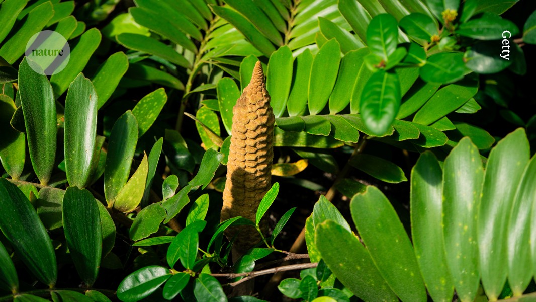
Exploring the Correlation Between the Serum Homocysteine to Apolipopro

Introduction

Coronary artery atherosclerotic cardiovascular disease (CAD) is a leading cause of mortality in developed countries. Common risk factors include hypercholesterolemia, hypertension, smoking, and diabetes, accounting for approximately 50% of all cases.1 Hyperhomocysteinemia (HHcy), occurring in 5% to 7% of the general population, is an important risk factor for coronary artery atherosclerotic heart disease.2 Studies have shown that elevated plasma homocysteine levels are associated with the formation of atherosclerosis, Coronary Artery Disease, acute myocardial infarction, arrhythmia formation, and sudden cardiac death.3–6 Possible mechanisms underlying the promotion of atherosclerosis include endothelial cell dysfunction, promotion of vascular smooth muscle cell (VSMC) proliferation, promotion of monocyte and leukocyte infiltration, stimulation of inflammatory cytokine expression, increased lipid peroxidation, and activation of thrombosis,7–11 among which lipid abnormalities are an important mechanism in the occurrence and development of CAD.

Apolipoprotein A-1 (ApoA-1), as the main protein component of HDL-C particles, also plays an important role in anti-atherosclerotic effects.12,13 ApoA-1 can exert its anti-atherosclerotic effects through cholesterol reverse transport and inhibition of inflammatory responses.13 However, homocysteine may inhibit the production of APOA1. Related cell experiments have shown that Hcy can reduce serum ApoA-1 levels by down-regulating the PPARα response element (PPRE) and reducing ApoA-1 promoter activity.14 Clinical studies have also found a negative correlation between plasma homocysteine and high-density lipoprotein cholesterol. A study on the effect of homocysteine on lipid metabolism in patients with CAD and non- CAD populations showed that the proportion of CAD patients increased with elevated plasma homocysteine levels, accompanied by decreased levels of APOA-1.15 This indicates that homocysteine plays an important role in lipid metabolism, so the ratio of homocysteine to APOA-1 (HAR) may better reflect the metabolic status of APOA-1 in patients with c CAD. Currently, there are no reports on the correlation between HAR and CAD. HAR may better reflect the imbalance of lipid metabolism in patients, to some extent reflecting the dysfunction of vascular endothelial function and inflammatory conditions in the body, which are indispensable factors in the occurrence and development of coronary artery atherosclerosis. Therefore, in this study, the Gensini score was used as the evaluation method for coronary artery lesions. By analyzing the correlation between HAR and the Gensini score, the evaluation value of HAR for coronary artery lesions was explored.

Materials and Methods

General Information

A retrospective analysis was conducted, and a total of 1596 inpatients admitted to the Cardiology Department of Xuan Cheng People’s Hospital due to chest pain from January 2019 to December 2021 were consecutively selected (experimental cohort). From January 2019 to December 2021, a total of 1468 inpatients admitted to the Cardiology Department of the 904th Hospital of the People’s Liberation Army Joint Logistics Support Force due to chest pain were consecutively selected (validation cohort). This study was approved by the Ethics Committee. Inclusion criteria: (1) Age 30–85 years, patients admitted due to chest pain scheduled for coronary angiography; (2) Patients with clear consciousness, able to communicate naturally, without serious neurological or psychiatric disorders. All subjects received standardized dual antiplatelet therapy with aspirin and ticagrelor/clopidogrel upon admission. According to the WHO guidelines for CAD diagnosis, stenosis ≥50% in any major coronary artery such as the left main, left anterior descending, left circumflex, right coronary artery, or its major branches (diagonal branch, obtuse marginal branch, left posterior descending artery, posterior descending branch) was used as the diagnostic criteria for coronary artery atherosclerotic heart disease (CAD). Exclusion criteria: (1) Patients with a history of old myocardial infarction, coronary artery intervention, or coronary artery bypass graft surgery; (2) Patients with acute cerebral infarction within the past six months; (3) Patients with pulmonary embolism, aortic dissection, acute or chronic nephritis, or other systemic diseases; (4) Patients with hematological diseases, malignant tumors, or autoimmune diseases; (5) Patients with acute or chronic infectious diseases; (6) Patients who did not undergo HCY or apolipoprotein A1 (ApoA-1) testing; (7) Patients who refused coronary angiography. Ultimately, 735 patients were included in the experimental cohort, including 264 in the control group, 51 in the coronary artery atherosclerosis group, and 420 in the CAD group. The validation cohort included 967 patients, including 220 in the control group, 170 in the coronary artery atherosclerosis group, and 577 in the CAD group. For parameters with missing data of less than 5%, the median of that parameter was used for imputation. All patients signed informed consent upon admission, agreeing to the use of their medical data for clinical research purposes.

Methods

Clinical data of all patients upon admission were collected, including gender, history of cerebral infarction, hypertension, diabetes, smoking, etc., indicated as present/absent. All patients had venous blood collected from the cubital vein on an empty stomach in the morning after admission for routine blood tests. Indicators such as apolipoprotein A1 (ApoA-1) and homocysteine (HCY) were measured using a fully automated biochemical analyzer (Beckman Coulter, USA), and the HAR (HCY/HDL-C) was calculated.

Grouping

According to coronary angiography and Gensini scores, all patients in the experimental cohort were divided into the control group (n=264), coronary artery atherosclerosis group (n=51), and CAD group (n=420), with the CAD group further divided into two subgroups: mild CAD subgroup (Gensini score <30, n=322) and severe coronary heart disease subgroup (Gensini score ≥30, n=98). Similarly, all patients in the validation cohort were divided into the control group (n=220), coronary artery atherosclerosis group (n=170), and CAD group (n=577), with the CAD group further divided into two subgroups: mild CAD subgroup (Gensini score <30, n=304) and severe CAD subgroup (Gensini score ≥30, n=273).

Statistical methods

Statistical analysis was performed using SPSS version 26.0 and R-studio statistical software (version 4.1.2). Quantitative data with normal distribution were expressed as mean ± standard deviation (Mean±SD), while data not conforming to normal distribution were expressed as median (interquartile range) [M (Q25, Q75)]. Between-group comparisons of non-normally distributed samples were performed using the two-sample Mann–Whitney U-test. Qualitative data were presented as counts and percentages, and between-group comparisons were conducted using the chi-square test. Performing univariate logistic regression and multivariate logistic regression analyses to identify independent factors associated with coronary artery lesions and severe coronary artery lesions. Spearman correlation analysis was used to evaluate the correlation between HAR and Gensini scores. Restricted cubic spline analysis was performed using the rms package (version 6.8–0) in R-studio statistical software to assess whether there was a linear relationship between HAR and CAD. Based on the results of multivariate logistic regression analysis, a nomogram was constructed using the rms package to calculate the C-index. The clinical application value and net benefit of HAR were evaluated using the decision curve analysis (DCA) with the rmad package (version 1.6).

Results

Baseline Data and Spline Analysis

The final experimental cohort included 735 patients, with 264 in the control group (133 males, 131 females), 51 in the coronary artery atherosclerosis group (31 males, 20 females), and 420 in the CAD group (254 males, 166 females). The validation cohort included 967 patients, with 220 in the control group (114 males, 106 females), 170 in the coronary artery atherosclerosis group (101 males, 69 females), and 577 in the CAD group (431 males, 146 females). Details of demographic and clinical characteristics are provided in Table 1.

Table 1 Demographics and Clinical Characteristics in the Derivation and External Validation Cohorts

In the experimental cohort, the relationship between HAR and CAD was analyzed using a restricted cubic spline model, Restricted cubic spline analysis in logistic regression revealed a sustained increase in the occurrence and development of CAD associated with HAR (Nonlinear P=0.436>0.05, Figure 1).

Figure 1 Restricted cubic spline plots for CAD by HAR levels. Restricted cubic spline plots were generated based on whether patients had coronary heart disease. The thick solid line represents the estimated adjusted odds ratio, with the light red shaded ribbon indicating the 95% confidence interval. The horizontal solid line represents an odds ratio of 1.0.

Comparison of General Information

General data comparison showed that the CAD group had older age, more male patients and smokers, a higher proportion of comorbidities such as hypertension and diabetes, decreased levels of serum HDL-C, ApoA-1, ALB, and total protein (P<0.05), and higher levels of homocysteine, monocytes (MONO), neutrophils (NE), white blood cells (WBC), and HAR compared to the control group (P<0.05, Table 2). Compared to the subgroup of mild CAD, there were more male patients, lower levels of APOA-1, ALB, and HDL-C (P<0.05), and significantly elevated levels of HCY, Scr, glucose, uric acid, monocytes, NE, WBC, and HAR (P<0.05, Table 3).

Table 2 Comparison of Clinical and Biochemical Data Between Normal Group and CAD

Table 3 Comparison of General Data Between Mild CAD Group and Severe CAD Group

CAD Risk Factors

Factors with statistically significant differences (P<0.05) between the CAD group and non-coronary artery group were included in a univariate logistic regression analysis to screen for factors affecting the occurrence of CAD. The results showed that age, gender, cerebral infarction, hypertension, diabetes, smoking, ALB, APOA-1, HCY, UA, Scr, MONO, NE, PLT, WBC, and HAR were predictive factors for CAD (P<0.05, Table 4).

Table 4 Univariate and Multivariate Logistic Regression Analysis of Influencing Factors of CAD

Factors with statistically significant differences (P<0.05) from the univariate logistic regression results were included in a multivariate logistic regression analysis. The collinearity diagnosis showed that the variance inflation factors of HCY and HAR were both greater than 10, and the tolerance was less than 0.1. Therefore, after removing HCY, factors such as age, gender, cerebral infarction, hypertension, diabetes, smoking, ALB, APOA-1, UA, Scr, MONO, NE, PLT, WBC, and HAR were included in the multiple-factor logistic regression analysis to construct a model for risk prediction. The results showed that age, hypertension, Diabetes, Smoking, WBC, and HAR were independent predictive factors for CAD (Table 4).

Construction of CAD Risk Prediction Model

Based on the results of multiple logistic regression analysis, we constructed three risk prediction models for CAD. Model 1: The risk prediction model was constructed based on HAR (Figure 2A), with a C-index of 0.634 (95% CI 0.595–0.674). Internal bootstrap validation was performed with repeated sampling (1,000 repetitions) to verify the nomogram model. The predictive model’s C-index was 0.635. Subsequently, model validation was conducted in the validation cohort, and based on HAR, a nomogram was constructed, yielding a predictive model’s C-index of 0.657 (Figure S1A). Model 2: A risk prediction model was built based on age, hypertension, diabetes, smoking, and WBC (Figure 2B), with a C-index of 0.692 (95% CI 0.654–0.730). Internal bootstrap validation was utilized with repeated sampling (1,000 repetitions) to verify the nomogram model, resulting in a predictive model’s C-index of 0.683. External validation yielded a predictive model’s C-index of 0.732 (Figure S1B). Model 3: A risk prediction model was established based on HAR combined with age, hypertension, diabetes, smoking, and WBC (Figure 2C), with a C-index of 0.719 (95% CI 0.682–0.756). Internal bootstrap validation was conducted with repeated sampling (1,000 repetitions) to verify the nomogram model, and the predictive model’s C-index was 0.710. External validation yielded a predictive model’s C-index of 0.750 (Figure S1C).

Figure 2 Nomogram for predicting the probability of CAD in Derivation cohort. (A) Model 1, The risk prediction model was constructed based on HAR. (B) Model 2: The risk prediction model was built based on age, hypertension, diabetes, smoking, and WBC. (C) Model 3: The risk prediction model was established based on HAR combined with age, hypertension, diabetes, smoking, and WBC.

Model performance evaluation: Goodness-of-fit analysis revealed that the calibration curves around the ideal model curve (the diagonal) exhibited slight fluctuations, indicating that the nomogram models had good calibration (Figure 3A–C). External validation: The nomogram models also demonstrated good calibration (Figure S2AS2C).

Figure 3 Calibration curve for predicting the probability nomogram of CAD in Derivation cohort. (A) Model 1 Calibration Curve; (B) Model 2 Calibration Curve; (C) Model 3 Calibration Curve; The calibration plot shows the agreement between the predicted (x-axis) and observed (y-axis) risks of CAD; the x-axis indicates the predicted risk of CAD, and the y-axis indicates the observed risk of CAD. Further, the calibration plot shows the predicted risk of in-hospital MACE for primary PCI after bootstrapping with 1000 replicates. The diagonal line indicates that the ideal curve is consistent with the actual curve and the solid line indicates the actual performance of the prediction model. The proximity of the solid line to the dashed diagonal line indicates the accuracy of the prediction model.

Factors Influencing the Severity of CAD

Univariate Logistic Regression Analysis of Factors Affecting CAD

Factors with statistically significant differences (P < 0.05) in general characteristics between the subgroups of mild and severe CAD were included in a univariate logistic regression analysis to screen for relevant factors influencing the severity of CAD. The results showed that age, gender, APOA-1, HDL, glucose, uric acid, MONO, WBC, and HAR were predictive factors for severe CAD (P < 0.05, Table 5).

Table 5 Univariate and Multivariate Logistic Regression Analysis of Severe CAD

Multivariable Logistic Regression Analysis of Factors Affecting the Severity of CAD

Factors with statistically significant differences (P < 0.05) in the univariate logistic regression analysis were included in the multivariate logistic regression analysis. Collinearity diagnostics suggested that the inflation factor of HCY and HAR exceeded 10, with tolerance less than 0.1. Therefore, after removing age, gender, APOA-1, HDL, glucose, uric acid, MONO, WBC, and HAR, these factors were included in the multivariate logistic regression analysis to construct the risk prediction model. The results showed that age [OR=1.033, 95% CI (1.008–1.059), P < 0.01], WBC [OR=1.250, 95% CI (1.101–1.420), P < 0.01], and HAR [OR=1.158, 95% CI (1.104–1.214), P < 0.01] were independent predictive factors for severe CAD (Table 5).

Construction of Risk Prediction Model for Severe Coronary Artery Lesions

Based on the results of the multivariate logistic regression analysis, a risk prediction model for severe CAD (Gensini score ≥30) was constructed. Model 1: The HAR-based risk prediction model (Figure 4A) had a C-index of 0.782 (95% CI 0.740–0.825). Internal bootstrap validation was used with repeated sampling (1,000 repetitions) to verify the nomogram model, with a predictive model C-index of 0.783. Model validation in the validation cohort yielded a predictive model C-index of 0.760 (Figure S3A). Model 2: A risk prediction model based on age, hypertension, diabetes, smoking, and WBC was constructed (Figure 4B), with a predictive model C-index of 0.697 (95% CI 0.641–0.754). Internal bootstrap validation was used with repeated sampling (1,000 repetitions) to verify the nomogram model, with a predictive model C-index of 0.694. External validation of the predictive model yielded a C-index of 0.721 (Figure S3B). Model 3: A risk prediction model combining HAR with age, hypertension, diabetes, smoking, and WBC was constructed (Figure 4C), with a predictive model C-index of 0.837 (95% CI 0.798–0.875). Internal bootstrap validation was used with repeated sampling (1,000 repetitions) to verify the nomogram model, yielding a predictive model C-index of 0.832. External validation of the predictive model yielded a C-index of 0.813 (Figure S3C).

Figure 4 Nomogram for predicting the probability of Severe CAD in Derivation cohort. (A) Model 1, The risk prediction model was constructed based on HAR. (B) Model 2: The risk prediction model was built based on age and WBC. (C) Model 3: The risk prediction model was established based on HAR combined with age and WBC.

Model performance evaluation of the nomogram: Goodness-of-fit analysis showed that the nomogram model exhibited good calibration (Figure 5A–C). External validation: The nomogram model also demonstrated good calibration (Figure S4AS4C).

Figure 5 Calibration curve for predicting the probability nomogram of Severe CAD in Derivation cohort. (A) Model 1 Calibration Curve; (B) Model 2 Calibration Curve; (C) Model 3 Calibration Curve.The calibration plot shows the agreement between the predicted (x-axis) and observed (y-axis) risks of CAD; the x-axis indicates the predicted risk of CAD, and the y-axis indicates the observed risk of CAD. Further, the calibration plot shows the predicted risk of in-hospital MACE for primary PCI after bootstrapping with 1000 replicates. The diagonal line indicates that the ideal curve is consistent with the actual curve and the solid line indicates the actual performance of the prediction model. The proximity of the solid line to the dashed diagonal line indicates the accuracy of the prediction model.

Correlation Between HAR and Gensini Score

The Spearman correlation analysis results showed a positive correlation between HAR and Gensini score (r=0.331, P<0.01). As HAR increased, the Gensini score also increased, indicating that higher HAR values were associated with more severe coronary artery lesions (Figure 6). External validation revealed similar results, with Spearman correlation analysis showing a positive correlation between HAR and Gensini score (r=0.393, P<0.01) (Figure S5).

Figure 6 Correlation Analysis of HAR and Gensini Score in Derivation cohort.

Assessment of DCA Curves for Evaluating HAR Clinical Performance

DCA compares the clinical performance of three nomogram models. Pt represents the threshold probability of severe CAD in the local population. DCA demonstrates that when Pt > 0, all three curve-line models start to achieve significant net benefit relative to the assumption of all patients having either negative or positive severe CAD. When Pt ranges between 0 and 0.89, compared to Model Two and Model One, Model Three incorporating HAR exhibits higher net benefit across a larger range of Pt, offering significant advantages in clinical practice (Figure 7). External validation results further indicate that Model Three maintains higher clinical net benefit over Model One and Model Two (Figure S6).

Figure 7 DCA curve analysis of the prediction model for severe coronary artery disease in Derivation cohort.

Comparative clinical performance of HAR with HCY, ApoA-1, WBC, and AGE in predicting severe CAD is assessed through DCA curves. Results suggest that when Pt ranges between 0 and 0.86, HAR demonstrates higher net benefit across a broader range of Pt compared to HCY, ApoA-1, WBC, and AGE, significantly outperforming these indicators (Figure 8). External validation results continue to indicate that HAR maintains higher clinical net benefit than other indicators (Figure S7).

Figure 8 Decision Curve Analysis of HAR and other parameters in Derivation cohort.

Discussion

ApoA-1, as the main protein component of HDL particles, plays a crucial role in the anti-atherosclerotic effect.16 ApoA-1 produced by the liver and intestine interacts with macrophage cholesterol transporter, initiating cholesterol efflux from macrophages, forming nascent HDL, which then transports cholesterol back to the liver in reverse cholesterol transport (RCT) via bile excretion.17 RCT effectively promotes cholesterol efflux from foam cells and limits the formation of atherosclerosis. ApoA-1 also clears oxidized phospholipids from oxLDL and cells. Studies have found that in preventing the oxidation of low-density lipoprotein, only reconstituted high-density lipoprotein (HDL) containing ApoA-1 and 1-palmitoyl-2-oleoyl phosphatidylcholine (POPC) is as effective as intact HDL, supporting the crucial antioxidative role of ApoA-1.18 Research by Chongren Tang et al found that ApoA-1 can inhibit stress by mediating cholesterol efflux through regulating lipid rafts.19 It can also inhibit the activation of p38 mitogen-activated protein kinase signaling induced by lipopolysaccharide, further promoting the generation of Foam cell zinc finger protein 36, thereby suppressing the release of related inflammatory factors.20 ApoA-1 can also block the activation of T lymphocytes contacting monocytes, inhibiting the production of inflammatory cytokines such as IL-6 and tumor necrosis factor-alpha.21

HCY is also an important risk factor in the occurrence and development of CAD. Studies have shown that HCY can promote inflammation, affect lipid metabolism, and thus cause coronary artery dysfunction, promoting the occurrence of CAD.22 High levels of HCY can also enhance the expression of the scavenger receptor CD36 in macrophages, leading to the formation of foam cells.23 Dan Liao et al24 found that high levels of HCY reduce circulating HDL-C by inhibiting the synthesis of ApoA-1 protein and promoting the clearance of HDL-C, inhibiting reverse cholesterol transport. HCY inhibits hepatic ApoA-1 expression through the peroxisome proliferator-activated receptor α (PPARα)-ApoA-1 pathway.14,25,26 Additionally, HCY can reduce ApoA-1 transcription by stimulating nuclear factor kappa B (NF-κB) and ApoA-1 regulatory protein-1 (ARP-1).27 Reduced plasma and hepatic expression levels of ApoA-1 have also been observed in animal models of hyperhomocysteinemia (HHcy).14,27 Artur Mierzecki et al also found that low-dose folic acid supplementation can reduce HCY concentrations and increase serum APOA1 concentration.28 The above studies all indicate a negative correlation between plasma HCY levels and APOA-1 in patients with CAD, and high levels of HCY can inhibit the generation of APOA-1, leading to a decrease in plasma HDL-C. This provides a theoretical basis for constructing the HCY-to-APOA-1 ratio (HAR).

There have been no reports on the combined exploration of the relationship between HCY and ApoA-1 in relation to CAD. We proposed the new index HAR for the first time based on the mechanism of interaction between HCY and APOA-1 in the body. Theoretically, HAR can to some extent reflect oxidative stress, inflammatory status, and lipid metabolism disorders in the body simultaneously. HCY is closely related to the occurrence and development of CAD, while ApoA-1 has antioxidative, anti-inflammatory, and anti-atherosclerotic effects and is significantly negatively correlated with the degree of coronary artery lesions.29 Therefore, the ratio of HCY to ApoA-1 may better reflect oxidative stress, inflammatory status, and lipid metabolism disorders in the course of CAD.

This study explored the correlation between HAR and other traditional inflammatory and lipid parameters with CAD and the severity of CAD. The results indicate that HAR has certain advantages in predicting CAD compared to other traditional inflammatory and lipid parameters. Restricted cubic spline analysis suggests that HAR is linearly correlated with the occurrence of CAD, and Spearman correlation analysis indicates that the more severe the coronary artery lesions, the higher the HAR levels. Multivariable logistic regression analysis supports that HAR is an independent risk factor for CAD and severe CAD. Subsequently, based on HAR, a Nomogram model was constructed. The C-index of the model indicates that the Nomogram model has good predictive value for CAD and severe CAD. External validation of the model also suggests that HAR has good predictive value for CAD and severe CAD. The calibration curve of the predictive model shows that the curve of the predictive model fluctuates slightly around the ideal model curve (diagonal line), indicating that the predictive model has good calibration, and there is good consistency between the predicted probability and the actual observation. DCA analysis suggests that HAR has good clinical utility.

Research Innovation

This study introduced a new risk parameter for CAD, namely the HAR, based on the interaction mechanism between HCY and ApoA-1. Utilizing data from two medical centers, the study investigated the relationship between HAR and the severity of CAD to enhance the accuracy and persuasiveness of the findings. The correlation between HAR and CAD severity was significantly stronger than that of ApoA-1 and HCY individually. Furthermore, the net clinical benefit of HAR in practice was markedly higher than that of ApoA-1 and HCY. As a novel risk factor for CAD, HAR demonstrates significant scientific, rational, and innovative value, contributing to the early diagnosis and intervention of CAD, thereby holding important clinical significance.

Limitations of the Study

1. The use of the Gensini score to evaluate CAD lacks features such as branch lesions, tortuous lesions, and calcified lesions, which may pose certain limitations. 2. HCY, ApoA-1, etc., were only measured as single preoperative blood samples without dynamic monitoring. The potential changes in HAR ratio after procedures such as percutaneous coronary intervention (PCI) or coronary artery bypass grafting (CABG) need further investigation. 3. This study only discusses the relationship between HAR and CAD, while its association with the long-term prognosis of CAD and the occurrence of adverse events requires further exploration. 4.In recent years, cardiac biomarkers such as high-sensitivity troponin T (hs-TnT) and N-terminal pro B-type natriuretic peptide (NT-proBNP) have become essential tools in the diagnosis, risk stratification, and prognosis of patients with coronary artery disease (CAD).30 However, in our study, hs-TnT and NT-proBNP measurements were not obtained for all patients, as these tests were selectively performed in high-risk individuals during pre-procedural assessment. Future studies should consider incorporating these markers systematically to further elucidate their relationship with HAR and CAD severity.

Conclusion

This study proposed a new CAD risk assessment parameter, namely the HAR, based on the interaction between HCY and ApoA-1. The investigation explored the relationship between HAR and the severity CAD, utilizing data from two medical centers to enhance accuracy and persuasiveness. As a novel risk factor for CAD, HAR holds significant scientific, rational, and innovative value. In clinical practice, HAR aids in the early diagnosis and intervention of CAD, thereby holding important clinical significance.

Data Sharing Statement

The datasets used and/or analyzed in this study are available from the corresponding authors upon reasonable request. Specifically, the validation cohort data is available from Shi Chen, and the experimental cohort data is available from Cunming Fang.

Ethics Approval and Consent to Participate

Ethical Statement: The authors are accountable for all aspects of the work in ensuring that questions related to the accuracy or integrity of any part of the work are appropriately investigated and resolved. The study was conducted in accordance with the Declaration of Helsinki (as revised in 2013). The study was approved by ethics committee of XuanCheng People’s Hospital (No. 2024-LW015-01). The study was approved by the medical ethics committee of The 904th Hospital of the PLA Joint Logistics Support Force. All methods were performed in accordance with the applicable guidelines and regulations. Informed consent was taken from all the patients.

Author Contributions

All authors made a significant contribution to the work reported, whether that is in the conception, study design, execution, acquisition of data, analysis and interpretation, or in all these areas; took part in drafting, revising or critically reviewing the article; gave final approval of the version to be published; have agreed on the journal to which the article has been submitted; and agree to be accountable for all aspects of the work. Jingshui Zhang and Zufei Wu contributed equally to the manuscript and should be considered as co-first authors. Shi Chen and CunMing Fang contributed equally to this work and should be considered as co-corresponding authors.

Funding

The authors declare that financial support was received for the research, authorship, and/or publication of this article. This study was supported by a University-Level Research grant from Wannan Medical College (Grant No. WK2023JXYY148).

Disclosure

The authors declare that there is no conflict of interest regarding the publication of this paper.

References

1. Yusuf S, Hawken S, Ounpuu S, et al. Effect of potentially modifiable risk factors associated with myocardial infarction in 52 countries (the INTERHEART study): case-control study. Lancet. 2004;364(9438):937–952. doi:10.1016/s0140-6736(04)17018-9

2. Hankey GJ, Eikelboom JW. Homocysteine and vascular disease. Lancet. 1999;354(9176):407–413. doi:10.1016/s0140-6736(98)11058-9

3. Zhang C-Y, Xu R-Q, Wang X-Q, et al. Comprehensive transcriptomics and metabolomics analyses reveal that hyperhomocysteinemia is a high risk factor for coronary artery disease in a Chinese obese population aged 40–65: a prospective cross-sectional study. Cardiovasc Diabetol. 2023;22(1):219. doi:10.1186/s12933-023-01942-0

4. Marković Boras M, Čaušević A, Brizić I, Mikulić I, Vasilj M, Jelić Knezović N. A relation of serum homocysteine, uric acid and C-reactive protein level in patients with acute myocardial infarction. Medicinski Glasnik. 2018;15(2):101–108. doi:10.17392/956-18

5. Rallidis LS, Kosmas N, Rallidi T, Pavlakis G, Kiouri E, Zolindaki M. Homocysteine is an independent predictor of long-term cardiac mortality in patients with stable coronary artery disease in the era of statins. Coron Artery Dis. 2020;31(2):152–156. doi:10.1097/mca.0000000000000800

6. Moshal KS, Metreveli N, Frank I, Tyagi SC. Mitochondrial MMP activation, dysfunction and arrhythmogenesis in hyperhomocysteinemia. Curr Vasc Pharmacol. 2008;6(2):84–92. doi:10.2174/157016108783955301

7. Lentz SR, Sobey CG, Piegors DJ, et al. Vascular dysfunction in monkeys with diet-induced hyperhomocyst(e)inemia. J Clin Invest. 1996;98(1):24–29. doi:10.1172/jci118771

8. Tsai JC, Wang H, Perrella MA, et al. Induction of cyclin A gene expression by homocysteine in vascular smooth muscle cells. J Clin Invest. 1996;97(1):146–153. doi:10.1172/jci118383

9. Wang H, Yoshizumi M, Lai K, et al. Inhibition of growth and p21 methylation in vascular endothelial cells by homocysteine but not cysteine. J Biol Chem. 1997;272(40):25380–25385. doi:10.1074/jbc.272.40.25380

10. Heinecke JW, Kawamura M, Suzuki L, Chait A. Oxidation of low density lipoprotein by thiols: superoxide-dependent and -independent mechanisms. J Lipid Res. 1993;34(12):2051–2061. doi:10.1016/S0022-2275(20)35347-5

11. Durand P, Lussier-Cacan S, Blache D. Acute methionine load-induced hyperhomocysteinemia enhances platelet aggregation, thromboxane biosynthesis, and macrophage-derived tissue factor activity in rats. FASEB j. 1997;11(13):1157–1168. doi:10.1096/fasebj.11.13.9367351

12. Liu M, Mei X, Herscovitz H, Atkinson D. N-terminal mutation of apoA-I and interaction with ABCA1 reveal mechanisms of nascent HDL biogenesis. J Lipid Res. 2019;60(1):44–57. doi:10.1194/jlr.M084376

13. Barrett TJ, Distel E, Murphy AJ, et al. Apolipoprotein AI promotes atherosclerosis regression in diabetic mice by suppressing myelopoiesis and plaque inflammation. Circulation. 2019;140(14):1170–1184. doi:10.1161/circulationaha.119.039476

14. Mikael LG, Genest J, Rozen R. Elevated homocysteine reduces apolipoprotein A-I expression in hyperhomocysteinemic mice and in males with coronary artery disease. Circ Res. 2006;98(4):564–571. doi:10.1161/01.RES.0000204825.66410.0b

15. Wu DF, Liao QC, Lu F, Wang Z, Yu K, Deng JL. Differential effects of hyperhomocysteinemia on the lipid profiles and lipid ratios between patients with and without coronary artery disease: a retrospective observational study. Medicine. 2022;101(52):e32464. doi:10.1097/md.0000000000032464

16. Chroni A, Kardassis D. HDL dysfunction caused by mutations in apoA-I and other genes that are critical for HDL biogenesis and remodeling. Curr Med Chem. 2019;26(9):1544–1575. doi:10.2174/0929867325666180313114950

17. Paul A, Lydic TA, Hogan R, Goo Y-H. Cholesterol acceptors regulate the lipidome of macrophage foam cells. Int J Mol Sci. 2019;20(15):3784. doi:10.3390/ijms20153784

18. Getz GS, Wool GD, Reardon CA. Apoprotein A-I mimetic peptides and their potential anti-atherogenic mechanisms of action. Curr Opin Lipidol. 2009;20(3):171–175. doi:10.1097/MOL.0b013e32832ac051

19. Tang C, Houston BA, Storey C, LeBoeuf RC. Both STAT3 activation and cholesterol efflux contribute to the anti-inflammatory effect of apoA-I/ABCA1 interaction in macrophages. J Lipid Res. 2016;57(5):848–857. doi:10.1194/jlr.M065797

20. Zhang M, Zhao G-J, Yin K, et al. Apolipoprotein A-1 binding protein inhibits inflammatory signaling pathways by binding to apolipoprotein A-1 in THP-1 macrophages. Circ J. 2018;82(5):1396–1404. doi:10.1253/circj.CJ-17-0877

21. Hyka N, Dayer J-M, Modoux C, et al. Apolipoprotein A-I inhibits the production of interleukin-1β and tumor necrosis factor-α by blocking contact-mediated activation of monocytes by T lymphocytes. Blood. 2001;97(8):2381–2389. doi:10.1182/blood.v97.8.2381

22. Khera AV, Kathiresan S. Genetics of coronary artery disease: discovery, biology and clinical translation. Nat Rev Genet. 2017;18(6):331–344. doi:10.1038/nrg.2016.160

23. Zanin RF, Bergamin LS, Morrone FB, Coutinho-Silva R, de Souza Wyse AT, Battastini AMO. Pathological concentrations of homocysteine increases IL-1β production in macrophages in a P2X7, NF-ĸB, and erk-dependent manner. Purinergic Signalling. 2015;11(4):463–470. doi:10.1007/s11302-015-9464-5

24. Liao D, Tan H, Hui R, et al. Hyperhomocysteinemia decreases circulating high-density lipoprotein by inhibiting apolipoprotein A-I protein synthesis and enhancing HDL cholesterol clearance. Circ Res. 2006;99(6):598–606. doi:10.1161/01.Res.0000242559.42077.22

25. Sood HS, Hunt MJ, Tyagi SC. Peroxisome proliferator ameliorates endothelial dysfunction in a murine model of hyperhomocysteinemia. Am J Physiol Lung Cell Mol Physiol. 2003;284(2):L333–341. doi:10.1152/ajplung.00183.2002

26. Xiao Y, Zhang Y, Lv X, et al. Relationship between lipid profiles and plasma total homocysteine, cysteine and the risk of coronary artery disease in coronary angiographic subjects. Lipids Health Dis. 2011;10(1):137. doi:10.1186/1476-511x-10-137

27. Morishima A, Ohkubo N, Maeda N, Miki T, Mitsuda N. NFκB regulates plasma apolipoprotein A-I and high density lipoprotein cholesterol through inhibition of peroxisome proliferator-activated receptor α. J Biol Chem. 2003;278(40):38188–38193. doi:10.1074/jbc.M306336200

28. Baszczuk A, Thielemann A, Musialik K, et al. The impact of supplementation with folic acid on homocysteine concentration and selected lipoprotein parameters in patients with primary hypertension. J Nutr Sci Vitaminol. 2017;63(2):96–103. doi:10.3177/jnsv.63.96

29. Tani S, Nagao K, Hirayama A. Association of systemic inflammation with the serum apolipoprotein A-1 level: a cross-sectional pilot study. J Cardiol. 2016;68(2):168–177. doi:10.1016/j.jjcc.2015.08.016

30. Duchnowski P, Śmigielski W. Usefulness of myocardial damage biomarkers in predicting cardiogenic shock in patients undergoing heart valve surgery. Kardiol Pol. 2024;82(4):423–426. doi:10.33963/v.phj.99553

Continue Reading