Sample Page
Volleyball Adds Transfer Štiglic – Northwestern Athletics
Written by
admin
in
4. Sports
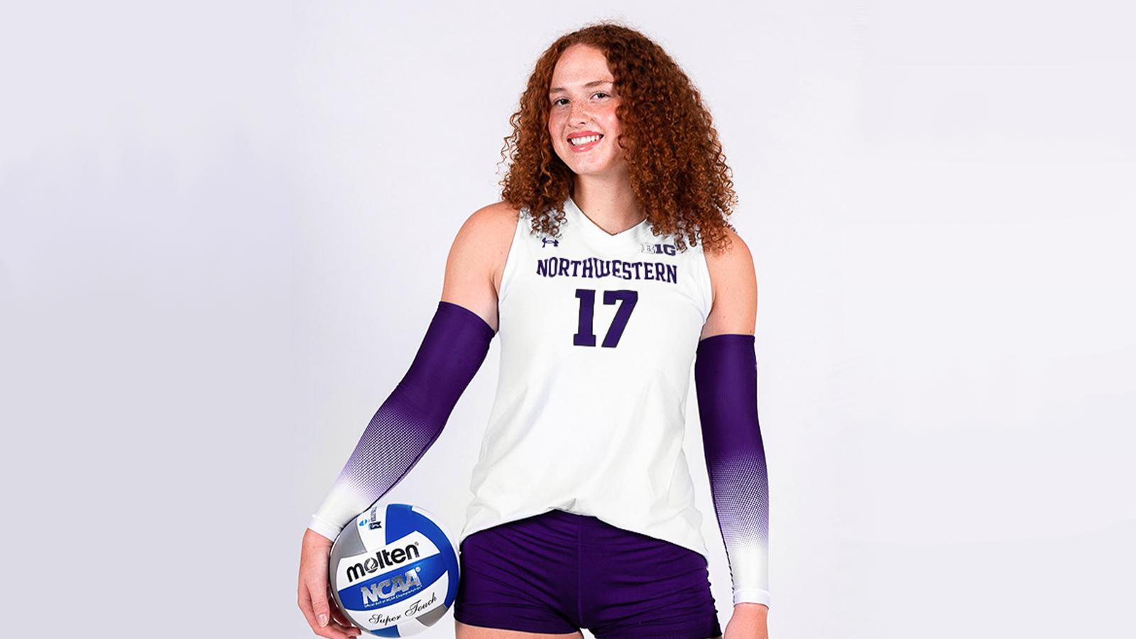
EVANSTON, Ill.
– Northwestern volleyball has added undergraduate transfer Mara Štiglic to the roster ahead of winter quarter, Head Coach
Tim Nollan
announced on Monday. Štiglic, a sophomore, will join the Wildcats after two seasons at Utah…
Continue Reading
←
Pakistan committed to empowering youth, creating transformative opportunities-Xinhua
The Solar System Loses an Ocean World
→
More posts
New Research Explores Paleolithic Transition from Neanderthals to Anatomically Modern Humans
December 22, 2025
Tariq Francis Named Co-Big Ten Player of the Week
December 22, 2025
Volleyball Ends Season Ranked 10th in Final AVCA Poll
December 22, 2025
Minnesota utility says it won’t buy from planned $1B power plant in Wisconsin
December 22, 2025