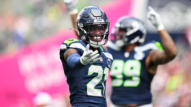
Introduction
Diffuse large B-cell lymphoma (DLBCL) is an aggressive B-cell lymphoma, the most common pathological type of NHL, accounting for approximately 30% to 40% of all NHL cases across different geographical regions.1,2 The median age at initial diagnosis of DLBCL is over 60 years, and 30% of patients are over 75 years old. The incidence of DLBCL increases with age.3,4 Epidemiological studies indicate that DLBCL has a complex and multifactorial etiology, including genetic characteristics, clinical features, and immune disorders, in addition to risk factors related to viruses, environment, high weight in youth, and occupational exposure.5,6 Although the prognostic significance of the International Prognostic Index (IPI) has been validated in many subtypes of NHL since 1993, its prognostic value in DLBCL remains controversial.
ABO blood group antigens, which play an important role in the physiology and pathology of cells, are defined by carbohydrate moieties on the extracellular surface of red blood cell membrane.7,8 Our previous research has elaborated on the relationship between ABO blood group and lymphoma, and summarized the current knowledge of the underlying pathogenic mechanisms of the association.9 It has been observed that ABO blood group is not only associated with the risk and prognosis of lymphoma, but may also be associated with the pathological classification of lymphoma patients.9 However, we did not specifically compare DLBCL with other lymphoma subtypes in our previous research. Given this background, we conducted a retrospective study specifically focusing on a representative pathological type, namely DLBCL, with the aim of investigating whether ABO blood group correlates with the risk of onset and prognosis of this disease. This study provides preliminary and exploratory evidence supporting ABO blood group as a potential biomarker for DLBCL. Its cost-effective and readily accessible nature warrants further validation in larger-scale studies, which may offer novel perspectives for future understanding of DLBCL-specific disease risk stratification and prognostic assessment.
Materials and Methods
We retrospectively analyzed 220 patients with newly diagnosed DLBCL at two medical institutions between January 2012 and December 2022. The research was conducted in full compliance with the guidelines set forth in the Declaration of Helsinki and obtained official authorization from the Institutional Review Board of the First Affiliated Hospital of Henan University of Science (No. 2024–1592 Fast). All patients with DLBCL participating in this study met the following inclusion criteria: (1) A diagnosis of DLBCL was confirmed by specialized pathologists according to the World Health Organization (WHO) classification. (2) No prior anti-cancer treatment had been administered. (3) Data on ABO blood group was accessible. (4) Sufficient clinical, laboratory, and follow-up records were available. Exclusion criteria include: (1) Transformed from other types of lymphoma to DLBCL. (2) Suffering from other tumors or having a history of tumor. (3) Suffering from other severe systemic diseases.
The baseline clinical data of patients were collected, including gender, age, Eastern Cooperative Oncology Group performance status (ECOG PS), primary tumor location, extranodal invasion details (sites and count), B symptoms, treatment modalities and response, ABO blood group, Ann Arbor stage, serum lactate dehydrogenase (LDH) levels, baseline serum CRP levels, serum β2-Microglobulin (β2-MG) levels, cellular origin, and IPI score. Overall survival (OS) is defined as the duration extending from the date of first diagnosis until either the occurrence of death from any cause or the last recorded date, when patient data is censored.
Additionally, we randomly selected age- and sex-matched hospitalized patients as controls (case-control ratio = 1) from the same institutions. Controls were diagnosed with non-malignant, non-hematological, and non-immunological disorders based on surgery or other routine clinical management (eg, hernia, cholelithiasis, osteoarthritis, cataract). Computerized randomization ensured equal numbers of controls per institution relative to DLBCL cases. ABO blood group data for controls were retrieved from hospital information systems (HIS) or laboratory databases using identical procedures as cases.
Within the DLBCL patient cohort, associations between ABO blood types and baseline clinical/laboratory variables were evaluated using Chi-square test or Fisher’s exact test for categorical data. When performing multiple pairwise comparisons among different blood groups for a specific variable, the Bonferroni correction was applied, adjusting the significance level to α’ = α / [k(k-1)/2], where k represented the number of blood groups, to account for all possible pairwise comparisons. The Log rank test and Kaplan-Meier method was applied for a univariate survival analysis. Variables demonstrating a univariate association with OS at P < 0.2 were included in multivariate Cox proportional hazards regression models. Hazard ratios (HRs) with 95% CIs were reported for significant predictors. A two-tailed P < 0.05 was deemed indicative of statistical significance. The statistical software package SPSS 26.0 (SPSS Inc., Chicago, IL, USA) was used for statistical calculations.
Result
Patient Characteristics
A total of 220 patients diagnosed with DLBCL, including 101 males and 119 females, with a median age of 60 years, were enrolled in the study. The clinical characteristics of the patients are listed in Table 1. Of the enrolled patients, 166 (75.5%) exhibited an optimal performance status (ECOG PS 0–1). B symptoms were present in 76 patients (34.5%). Involvement of at least two extranodal sites was displayed by 81 patients (36.8%). Elevated LDH levels were observed in 111 (50.5%) patients. The serum CRP levels were available for 108 patients, and the serum β2-MG data were available for 158 patients. Localized disease (stage I/II) was observed in 73 patients (33.2%). High-risk disease (IPI ≥ 3) was present in 79 patients (35.9%). Ki-67 antigen levels were available for 195 patients. Among the 220 patients with DLBCL, 115 (73.2%) originated from the non-germinal center B cell-like (GCB) subtype. The ABO blood group exhibited no significant association with patient age, gender, ECOG PS, B symptoms, the number of extranodal sites, LDH levels, CRP levels, serum β2-MG levels, Ann Arbor stage, IPI score, Ki-67 levels, or cellular origin (all P > 0.05, Table 1).
Table 1 Basic Characteristics of DLBCL Patients in Distinct ABO Blood Type Groups
|
The Effect of ABO Blood Group on Risk of DLBCL
In the DLBCL cohort, the distribution of ABO blood types was as follows: blood type A in 66 patients (30.0%), blood type B in 56 patients (25.5%), blood type AB in 24 patients (10.9%), and blood type O in 74 patients (33.6%). A control group comprising 220 individuals with nonmalignant conditions was randomly selected for comparison. The distribution of ABO blood types within the control group was as follows: blood group A accounted for 65 patients (29.5%), blood group B accounted for 72 patients (32.8%), blood group AB accounted for 17 patients (7.7%), and blood group O accounted for 66 patients (30.0%). No statistically significant disparity was observed in the distribution of ABO blood groups between DLBCL patients and the control cohort (P = 0.301, Supplementary Table 1).
Upon conducting a gender-stratified comparative analysis, we identified a statistically significant disparity among female patients with DLBCL compared to the control group (P = 0.012, Figure 1). Conversely, an analysis of the ABO blood group distribution among male DLBCL patients relative to the control group revealed no statistically significant differences (P = 0.757, Figure 1).
![]() |
Figure 1 Distribution of ABO blood types among DLBCL patients and controls by gender. Significant difference observed in females (P = 0.012, chi-square test); no significant difference observed in males (P = 0.757, chi-square test). Abbreviation: DLBCL, diffuse large B cell lymphoma.
|
In the study comparing female patients with DLBCL to a female control group without the disease, the prevalence rate of DLBCL were observed to be 54.5%, 34.3%, 70.0%, and 54.1% respectively in individuals with blood type A, B, AB, and O. To account for multiple pairwise comparisons across blood groups, Bonferroni correction was applied, yielding an adjusted significance threshold of α = 0.05/[4(4−1)/2] = 0.0083. Subsequent pairwise analysis demonstrated a significantly lower DLBCL risk in individuals with blood type B compared to blood type AB (P = 0.005, Table 2). No statistically significant differences in DLBCL risk were observed between other blood group pairs (P > 0.0083, Table 2).
![]() |
Table 2 DLBCL and the Distribution of ABO Blood Groups in Females
|
The Effect of ABO Blood Group on Survival of Patients with DLBCL
By the conclusion of the final follow-up period, a cumulative total of 77 (35.0%) patients had unfortunately passed away. The deaths were due to tumor progression (n = 69), severe pulmonary infections (n = 5), cardiovascular disease (n = 1), and other causes (n = 2). The 3-year OS rates for blood type A, B, AB, and O groups were 51.0%, 58.8%, 74.9%, and 74.0%, respectively (P = 0.458, Figure 2). Upon stratifying by age groups, we observed that among patients with DLBCL aged over 60 years, the 3-year OS rates for blood type A, B, AB, and O groups were 32.0%, 23.7%, 87.5%, and 69.0%, respectively, yielding a statistically significant difference (P = 0.043, Figure 3a). Considering that DLBCL patients with blood type B had the shortest 3-year OS rate, we categorized those aged over 60 into two distinct groups: blood type B and non-B (A, AB, and O). Patients with blood type B demonstrated a significantly reduced 3-year OS rate compared to those with non-B blood types (23.7% vs 53.6%, P = 0.030, Figure 3b). In contrast, among DLBCL patients aged 60 years or younger, no significant difference in survival rates was observed between individuals with blood type B and those with non-B blood types, with 3-year OS rates of 83.3% and 73.7%, respectively (P = 0.196, Figure 3c). Given that the 3-year OS rates of patients aged over 60 years with A and B blood types were shorter than those with AB and O blood types, we conducted a further comparison between blood type AB/O and blood type A/B to investigate the impact of ABO blood type on survival outcomes. The analysis revealed that the OS for patients with A/B blood types was significantly shorter compared to those with AB/O blood types (P = 0.014, Figure 3d). Notably, the 106 DLBCL patients aged over 60 years shared a similar clinical background (all P > 0.05, Supplementary Table 2).
![]() |
Figure 2 The Kaplan-Meier curves for OS in patients with DLBCL according to ABO blood type (P = 0.458 by Log rank test). Abbreviation: OS, overall survival; DLBCL, diffuse large B-cell lymphoma; A, blood type A; B, blood type B; AB, blood type AB; O, blood type O.
|
![]() |
Figure 3 The Kaplan-Meier curves for OS in patients with DLBCL according to ABO blood type. (a): OS in patients aged >60 years stratified by blood types A, B, AB, and O c. (b): OS in patients aged >60 years comparing blood type B vs non-B (A, O, and AB) (P = 0.030 by Log rank test). (c): OS in patients aged ≤60 years comparing blood type B vs non-B (A, O, and AB) (P = 0.196 by Log rank test). (d): OS in patients aged >60 years comparing blood types A/B vs AB/O (P = 0.014 by Log rank test). Abbreviation: OS, overall survival; DLBCL, diffuse large B-cell lymphoma; A, blood type A; B, blood type B; AB, blood type AB; O, blood type O; A/B, blood type A and blood type B; AB/O, blood type AB and blood type O.
|
Univariate and Multivariate Cox Regression Analysis
Table 3 presented the findings from both univariate and multivariate regression analyses regarding potential predictors of OS in patients with DLBCL aged over 60 years. The univariate analysis indicated that Ann Arbor stage, LDH levels, IPI score, and ABO blood type were significant prognostic factors influencing OS in patients with DLBCL (P < 0.05). Blood type B was linked to a significantly shorter OS when compared to non-B blood types (HR 2.013, 95% CI 1.056–3.839, P = 0.034). In the multivariate analysis, IPI score ≥ 3 (HR 2.247, 95% CI 1.226–4.120, P = 0.009), elevated LDH levels (HR 1.890, 95% CI 1.015–3.520, P = 0.045), and blood type B (HR 2.050, 95% CI 1.069–3.933, P = 0.031) emerged as adverse factors for OS.
![]() |
Table 3 Univariate and Multivariate Analysis of Prognostic Factors for OS in DLBCL Patients Aged Over 60 years
|
Discussion
In the present study, we found that females with blood type B might exhibit a reduced risk of DLBCL compared to those with blood type AB. The prognostic implications of ABO blood group distinctions were not apparent across the entire cohort of DLBCL patients. However, our analysis found notable prognostic significance associated with ABO blood group specifically among DLBCL patients aged over 60 years. Among these patients, those with blood type B experienced a significantly shorter OS compared to patients with non-B blood groups.
The ABO gene is located on chromosome 9q34 and encodes two alleles (ie, A and B) for specific glycosyltransferases that catalyze the covalent linkage of N-acetyl-D-galactosamine or D-galactose to a common precursor side chain (ie, the H antigen), eventually forming A and B antigens respectively.10,11 Unlike the A and B alleles, the O variant encodes a non-functional glycosyltransferase, so the H antigen remains unmodified.12 In recent years, researchers have found a possible association between ABO blood group and the development of cancers. Studies have indicated that individuals with blood type A may be at an increased risk of tumorigenesis, whereas those with blood type B appear to have a reduced risk.13–17 Previous investigations did not observe statistically significant results regarding the correlation between ABO blood group and the risk of DLBCL.18,19 This study provided evidence that among female patients, individuals with blood type B may have exhibited a decreased risk of developing DLBCL in comparison to those with AB blood types.
Epidemiological studies have shown that the incidence of DLBCL is significantly higher among males compared to females.20 This disparity may be linked to the presence of estrogen in the female population. Studies propose that estrogen potentially exhibits antitumor properties, capable of inhibiting the proliferation and dissemination of tumor cells through a variety of mechanisms.21 It has been reported that the use of high-dose oral contraceptives for pregnancy prevention or exposure to estrogen via postmenopausal hormone replacement therapy may reduce the risk of aggressive lymphoma.22 Furthermore, B-cell lymphomas treated with estrogen receptor β were shown to have effectively inhibit tumor growth in vivo.23 These findings provided additional evidence that estrogen played a significant role in the development and progression of lymphoma. The study suggested that, compared to females with blood type AB, those with blood type B might exhibit a reduced risk of developing DLBCL. The study suggested that, compared to females with blood type AB, those with blood type B might exhibit a reduced risk of developing DLBCL. We hypothesize that this may be partially mediated by the higher estrogen levels typically found in individuals with blood type B, though this remains speculative in the absence of direct hormonal measurements. Further research is warranted to substantiate this hypothesis.
There were few studies exploring the prognostic relationship between ABO blood groups and DLBCL, and the results were inconsistent. A study in Turkey revealed that there was no significant correlation between ABO blood groups and the prognosis of patients with DLBCL.19 This finding was consistent with the result of this study conducted among the entire cohort of DLBCL patients. Nevertheless, what distinguished it was that our subgroup analysis identified blood type B as a negative prognostic factor specifically for patients older than 60 years. Osada et al reported that DLBCL patients with blood type B had a shorter OS than those with non-B blood types, and this trend was more significant among male DLBCL patients.18 A large-scale, population-based study on DLBCL series showed that male patients had worse prognosis outcomes than female patients.24 Although our study observed similar results in DLBCL patients aged over 60 years, we did not find any relationship between gender and the survival of DLBCL patients.
The underlying mechanisms of how the ABO blood group may interact with the development and progression of cancers, including lymphoma, are still poorly understood. Several plausible hypotheses have been formulated to elucidate the link between ABO blood group and cancer risk. It is hypothesized that the absence of blood group antigen expression – particularly A and B antigens – may enhance tumor malignancy by increasing cellular motility and migration, thereby correlating with adverse clinical outcomes and poorer overall prognosis.25–27 Studies have indicated that the reduction or absence of ABO blood group antigen expression might be related to the deletion of ABO allele or relative down-regulation of the glycosyltransferase necessary for blood group antigen synthesis caused by hypermethylation of the ABO promoter region.28–32 The absence of ABO blood group antigens has been observed in hematological malignancies, including Hodgkin’s lymphoma (HL).33,34 We hypothesize that analogous mechanisms may be present in patients aged over 60 years with DLBCL, which could lead to the reduction or absence of B-type antigens, ultimately resulting in unfavorable prognostic outcomes. The glycosylation of ABO blood group antigens can lead to conformational changes in proteins that not only affect intercellular signaling, cell adhesion, and immune surveillance, but also stimulate tumor growth and metastasis.35–40 Some studies have reported that the ABO gene locus is associated with circulating levels of tumor necrosis factor-alpha, soluble intercellular adhesion molecule (ICAM)-1, E-selectin, and P-selectin.41–43 These adhesion molecules play a crucial role in the recruitment processes associated with chronic inflammation. Chronic inflammation is linked to tumor growth, invasion, and migration.44–46 Chronic inflammation is also associated with lymphatic malignancies.47 For example, the lymphomas that appear in mice deficient in GM-CSF and IFNγ are caused by infections and subside after antibiotic treatment.48 Although this study did not find a significant association between ABO blood group antigens and CRP, there may be other inflammatory cytokines that serve as intermediaries linking ABO blood group antigens to DLBCL. It is possible that ABO blood group antigens influence tumor progression and metastasis by altering the inflammatory state of the host. ABO glycosyltransferase can regulate plasma von Willebrand factor (vWF) levels, affecting the risk of venous thromboembolism.49,50 vWF plays an important role in inhibiting angiogenesis, promoting wound healing, and inducing tumor cell apoptosis; particularly, angiogenesis and apoptosis are also involved in tumorigenesis.51–54 Therefore, ABO blood group may contribute to the development of tumors by regulating plasma vWF levels.9 In this study, we observed a case of patients with DLBCL and blood type B who died from a pulmonary embolism. We observed one blood type B patient dying from pulmonary embolism, suggesting thromboembolic events as another potential mechanism.
This study has several limitations. First, Retrospective design inherently restricts causal inference and may introduce unmeasured confounders. Second, Absence of data on estrogen levels precludes validation of the proposed biological hypotheses. Third, the relatively small sample size with regionally constrained recruitment limits population-level generalizability and increases susceptibility to selection bias. Last, reduced statistical power after Bonferroni correction for multiple comparisons may have obscured subtle associations between other blood group.
Conclusion
In summary, our research found that females with blood type B may have a lower risk of developing DLBCL compared to females with blood type AB. Furthermore, blood type B may serve as a poor prognostic factor for patients over the age of 60 who have DLBCL. To better understand the role of ABO blood groups in DLBCL, future studies are recommended in a large number of different populations (Asian, Caucasian, African) as well as in various regions.
Data Sharing Statement
The original contributions presented in the study are included in the article/supplementary material. Further inquiries can be directed to the corresponding authors.
Ethics Approval and Consent to Participate
The studies involving humans were approved by the ethics committee of The First Affiliated Hospital of Henan University of Science and Technology. The studies were conducted in accordance with the local legislation and institutional requirements. All participants confirmed their informed consent by responding to yes/no inquiries. All information collected from this study was treated with utmost confidentiality.
Author Contributions
All authors made a significant contribution to the work reported, whether that is in the conception, study design, execution, acquisition of data, analysis and interpretation, or in all these areas; took part in drafting, revising or critically reviewing the article; gave final approval of the version to be published; have agreed on the journal to which the article has been submitted; and agree to be accountable for all aspects of the work.
Funding
This work was supported by the Doctoral Research Funds of Henan University of Science and Technology.
Disclosure
The authors report no conflicts of interest in this work.
References
1. Swerdlow SH, Campo E, Pileri SA, et al. The 2016 revision of the world health organization classification of lymphoid neoplasms. Blood. 2016;127(20):2375–2390. doi:10.1182/blood-2016-01-643569
2. Armitage JO, Gascoyne RD, Lunning MA, Cavalli F. Non-Hodgkin lymphoma. Lancet. 2017;390(10091):298–310. doi:10.1016/S0140-6736(16)32407-2
3. Sehn LH, Salles G. Diffuse large b-cell lymphoma. N Engl J Med. 2021;384(9):842–858. doi:10.1056/NEJMra2027612
4. Liu Y, Barta SK. Diffuse large B-cell lymphoma: 2019 update on diagnosis, risk stratification, and treatment. Am J Hematol. 2019;94(5):604–616. doi:10.1002/ajh.25460
5. Cerhan JR, Kricker A, Paltiel O, et al. Medical history, lifestyle, family history, and occupational risk factors for diffuse large B-cell lymphoma: the interlymph non-hodgkin lymphoma subtypes project. J Natl Cancer Inst Monogr. 2014;2014(48):15–25. doi:10.1093/jncimonographs/lgu010
6. De Roos AJ, Schinasi LH, Miligi L, et al. Occupational insecticide exposure and risk of non-hodgkin lymphoma: a pooled case-control study from the interlymph consortium. Int J Cancer. 2021;149(10):1768–1786. doi:10.1002/ijc.33740
7. Mohandas N, Narla A. Blood group antigens in health and disease. Curr Opin Hematol. 2005;12(2):135–140. doi:10.1097/01.moh.0000153000.09585.79
8. Storry JR, Olsson ML. The ABO blood group system revisited: a review and update. Immunohematology. 2009;25(2):48–59. doi:10.21307/immunohematology-2019-231
9. Qin L, Gao D, Wang Q, et al. ABO blood group and the risk and prognosis of lymphoma. J Inflamm Res. 2023;16:769–778. doi:10.2147/JIR.S401818
10. Yamamoto F. Molecular genetics of ABO. Vox Sang. 2000;78(2):91–103. doi:10.1111/j.1423-0410.2000.tb00045.x
11. Yamamoto F, Cid E, Yamamoto M, Blancher A. ABO research in the modern era of genomics. Transfus Med Rev. 2012;26(2):103–118. doi:10.1016/j.tmrv.2011.08.002
12. Lowe JB. The blood group-specific human glycosyltransferases. Baillieres Clin Haematol. 1993;6(2):465–492. doi:10.1016/s0950-3536(05)80155-6
13. Poole EM, Gates MA, High BA, et al. ABO blood group and risk of epithelial ovarian cancer within the Ovarian Cancer Association Consortium. Cancer Causes Control. 2012;23(11):1805–1810. doi:10.1007/s10552-012-0059-y
14. Sheng L, Sun X, Zhang L, Su D. ABO blood group and nasopharyngeal carcinoma risk in a population of Southeast China. Int J Cancer. 2013;133(4):893–897. doi:10.1002/ijc.28087
15. Li X, Xu H, Ding Z, Jin Q, Gao P. Association between ABO blood group and HCV-related hepatocellular carcinoma risk in China. Medicine (Baltimore). 2016;95(49):e5587. doi:10.1097/MD.0000000000005587
16. Li X, Xu H, Gao P. ABO blood group and diabetes mellitus influence the risk for pancreatic cancer in a population from China. Med Sci Monit. 2018;24:9392–9398. doi:10.12659/MSM.913769
17. Huang JY, Wang R, Gao YT, Yuan JM. ABO blood type and the risk of cancer – Findings from the Shanghai Cohort Study. PLoS One. 2017;12(9):e0184295. doi:10.1371/journal.pone.0184295
18. Osada Y, Ito C, Nishiyama-Fujita Y, et al. Prognostic impact of ABO blood group on survival in patients with malignant lymphoma. Clin Lymphoma Myeloma Leuk. 2020;20(2):122–129. doi:10.1016/j.clml.2019.09.607
19. Ulu BU, Başcı S, Bakırtaş M, et al. Could blood groups have prognostic significance on survival in patients with diffuse large B cell lymphoma. Leuk Res. 2022;115:106810. doi:10.1016/j.leukres.2022.106810
20. Morton LM, Wang SS, Devesa SS, Hartge P, Weisenburger DD, Linet MS. Lymphoma incidence patterns by WHO subtype in the United States, 1992-2001. Blood. 2006;107(1):265–276. doi:10.1182/blood-2005-06-2508
21. Pierdominici M, Maselli A, Locatelli SL, et al. Estrogen receptor β ligation inhibits Hodgkin lymphoma growth by inducing autophagy. Oncotarget. 2017;8(5):8522–8535. doi:10.18632/oncotarget.14338
22. Lee JS, Bracci PM, Holly EA. Non-Hodgkin lymphoma in women: reproductive factors and exogenous hormone use. Am J Epidemiol. 2008;168(3):278–288. doi:10.1093/aje/kwn119
23. Yakimchuk K, Hasni MS, Guan J, Chao MP, Sander B, Okret S. Inhibition of lymphoma vascularization and dissemination by estrogen receptor β agonists. Blood. 2014;123(13):2054–2061. doi:10.1182/blood-2013-07-517292
24. Székely E, Hagberg O, Arnljots K, Jerkeman M. Improvement in survival of diffuse large B-cell lymphoma in relation to age, gender, international prognostic index and extranodal presentation: a population based swedish lymphoma registry study. Leuk Lymphoma. 2014;55(8):1838–1843. doi:10.3109/10428194.2013.853297
25. Hakomori S. Antigen structure and genetic basis of histo-blood groups A, B and O: their changes associated with human cancer. Biochim Biophys Acta. 1999;1473(1):247–266. doi:10.1016/s0304-4165(99)00183-x
26. Le Pendu J, Marionneau S, Cailleau-Thomas A, Rocher J, Le Moullac-Vaidye B, Clément M. ABH and Lewis histo-blood group antigens in cancer. APMIS. 2001;109(1):9–31. doi:10.1111/j.1600-0463.2001.tb00011.x
27. Dabelsteen E, Gao S. ABO blood-group antigens in oral cancer. J Dent Res. 2005;84(1):21–28. doi:10.1177/154405910508400103
28. Stellner K, Hakomori S, Warner GS. Enzymic conversion of “H1-glycolipid” to A or B-glycolipid and deficiency of these enzyme activities in adenocarcinoma. Biochem Biophys Res Commun. 1973;55(2):439–445. doi:10.1016/0006-291x(73)91106-6
29. Orlow I, Lacombe L, Pellicer I, et al. Genotypic and phenotypic characterization of the histoblood group ABO(H) in primary bladder tumors. Int J Cancer. 1998;75(6):819–824. doi:10.1002/(sici)1097-0215(19980316)75:6<819::aid-ijc1>3.0.co;2-y
30. Iwamoto S, Withers DA, Handa K, Hakomori S. Deletion of A-antigen in a human cancer cell line is associated with reduced promoter activity of CBF/NF-Y binding region, and possibly with enhanced DNA methylation of A transferase promoter. Glycoconj J. 1999;16(10):659–666. doi:10.1023/a:1007085202379
31. Kominato Y, Hata Y, Takizawa H, Tsuchiya T, Tsukada J, Yamamoto F. Expression of human histo-blood group ABO genes is dependent upon DNA methylation of the promoter region. J Biol Chem. 1999;274(52):37240–37250. doi:10.1074/jbc.274.52.37240
32. Gao S, Bennett EP, Reibel J, et al. Histo-blood group ABO antigen in oral potentially malignant lesions and squamous cell carcinoma–genotypic and phenotypic characterization. APMIS. 2004;112(1):11–20. doi:10.1111/j.1600-0463.2004.apm1120103.x
33. Scott GL, Rasbridge MR. Loss of blood group antigenicity in a patient with Hodgkin’s disease. Vox Sang. 1972;23(5):458–460. doi:10.1111/j.1423-0410.1972.tb03836.x
34. Bianco T, Farmer BJ, Sage RE, Dobrovic A. Loss of red cell A, B, and H antigens is frequent in myeloid malignancies. Blood. 2001;97(11):3633–3639. doi:10.1182/blood.v97.11.3633
35. Greenwell P. Blood group antigens: molecules seeking a function. Glycoconj J. 1997;14(2):159–173. doi:10.1023/a:1018581503164
36. Pinho SS, Reis CA. Glycosylation in cancer: mechanisms and clinical implications. Nat Rev Cancer. 2015;15(9):540–555. doi:10.1038/nrc3982
37. Stowell SR, Ju T, Cummings RD. Protein glycosylation in cancer. Annu Rev Pathol. 2015;10:473–510. doi:10.1146/annurev-pathol-012414-040438
38. Xu Y, Chang R, Xu F, et al. N-glycosylation at asn 402 stabilizes n-cadherin and promotes cell-cell adhesion of glioma cells. J Cell Biochem. 2017;118(6):1423–1431. doi:10.1002/jcb.25801
39. Läubli H, Borsig L. Altered cell adhesion and glycosylation promote cancer immune suppression and metastasis. Front Immunol. 2019;10:2120. doi:10.3389/fimmu.2019.02120
40. Reily C, Stewart TJ, Renfrow MB, Novak J. Glycosylation in health and disease. Nat Rev Nephrol. 2019;15(6):346–366. doi:10.1038/s41581-019-0129-4
41. Melzer D, Perry JR, Hernandez D, et al. A genome-wide association study identifies protein quantitative trait loci (pQTLs). PLoS Genet. 2008;4(5):e1000072. doi:10.1371/journal.pgen.1000072
42. Kiechl S, Paré G, Barbalic M, et al. Association of variation at the ABO locus with circulating levels of soluble intercellular adhesion molecule-1, soluble P-selectin, and soluble E-selectin: a meta-analysis. Circ Cardiovasc Genet. 2011;4(6):681–686. doi:10.1161/CIRCGENETICS.111.960682
43. Barbalic M, Dupuis J, Dehghan A, et al. Large-scale genomic studies reveal central role of ABO in sP-selectin and sICAM-1 levels. Hum Mol Genet. 2010;19(9):1863–1872. doi:10.1093/hmg/ddq061
44. Fernandes JV, Cobucci RN, Jatobá CA, et al. The role of the mediators of inflammation in cancer development. Pathol Oncol Res. 2015;21(3):527–534. doi:10.1007/s12253-015-9913-z
45. Singh R, Mishra MK, Aggarwal H. Inflammation, Immunity, and Cancer. Mediators Inflamm. 2017;2017:6027305. doi:10.1155/2017/6027305
46. Greten FR, Grivennikov SI. Inflammation and cancer: triggers, mechanisms, and consequences. Immunity. 2019;51(1):27–41. doi:10.1016/j.immuni.2019.06.025
47. Grivennikov SI, Greten FR, Karin M. Immunity, inflammation, and cancer. Cell. 2010;140(6):883–899. doi:10.1016/j.cell.2010.01.025
48. Enzler T, Gillessen S, Manis JP, et al. Deficiencies of GM-CSF and interferon gamma link inflammation and cancer. J Exp Med. 2003;197(9):1213–1219. doi:10.1084/jem.20021258
49. Ibrahim-Kosta M, Bailly P, Silvy M, et al. ABO blood group, glycosyltransferase activity and risk of venous thromboembolism. Thromb Res. 2020;193:31–35. doi:10.1016/j.thromres.2020.05.051
50. Ward SE, O’Sullivan JM, O’Donnell JS. The relationship between ABO blood group, von Willebrand factor, and primary hemostasis. Blood. 2020;136(25):2864–2874. doi:10.1182/blood.2020005843
51. Starke RD, Ferraro F, Paschalaki KE, et al. Endothelial von Willebrand factor regulates angiogenesis. Blood. 2011;117(3):1071–1080. doi:10.1182/blood-2010-01-264507
52. Franchini M, Frattini F, Crestani S, Bonfanti C, Lippi G. von Willebrand factor and cancer: a renewed interest. Thromb Res. 2013;131(4):290–292. doi:10.1016/j.thromres.2013.01.015
53. O’Sullivan JM, Preston R, Robson T, O’Donnell JS. Emerging roles for von willebrand factor in cancer cell biology. Semin Thromb Hemost. 2018;44(2):159–166. doi:10.1055/s-0037-1607352
54. Ishihara J, Ishihara A, Starke RD, et al. The heparin binding domain of von Willebrand factosr binds to growth factors and promotes angiogenesis in wound healing. Blood. 2019;133(24):2559–2569. doi:10.1182/blood.2019000510