Unlock the Editor’s Digest for free
Roula Khalaf, Editor of the FT, selects her favourite stories in this weekly newsletter.
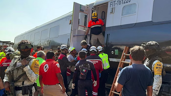
A train careered off the tracks in southern Mexico on Sunday, killing 13 people and injuring dozens more, in an incident…
Unlock the Editor’s Digest for free
Roula Khalaf, Editor of the FT, selects her favourite stories in this weekly newsletter.
A train careered off the tracks in southern Mexico on Sunday, killing 13 people and injuring dozens more, in an incident…