The winter season is upon us, and with it come chilly gusts of wind and the long-awaited winter holidays. Pakistan’s airports are entering one of their busiest periods of the year.
From early-morning departures to late-night arrivals, travellers are navigating tight schedules, heavy luggage, and the hassle of finding the right bag at the conveyor belt, while also braving the unpredictable traffic situation, which can go from bad to worse in a matter of minutes, making the journey to and from the airport just as critical and high-stakes as the flight itself.
During the last few years, it has been noticed how airport mobility has become a major concern for urban commuters. This has resulted in a rise in missed flights due to traffic delays, cancellations by traditional transport at the very last minute, and uncertainty around arrival times. All of these factors have made travellers increasingly cautious, and understandably so.
That is why more passengers are turning to ride-hailing platforms, as they offer predictable travel times, transparent pricing, and the option of real-time tracking. This is where platforms like Yango step in because they are increasingly being considered by travellers who want more control over timing, pricing, and predictability during airport commutes.
Industry observers note that winter travel is attached to its own set of challenges. Some of these include foggy mornings, reduced visibility, and seasonal congestion. These often end up disrupting road traffic, while international and domestic flights continue to operate on strict schedules.
For passengers travelling with families or carrying heavy luggage, it’s not just about the cost anymore. Reliability takes first place in such situations in order to avoid any unpleasant situations. For many travellers navigating these winter challenges, ride-hailing services such as Yango Ride offer a more reliable alternative during time-sensitive airport journeys.
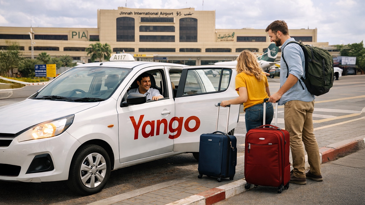
Yango Ride has positioned itself as a dependable option for getting to and from the airport during busy travel spells. The platform has seen increased adoption during peak travel periods, particularly among passengers prioritising punctuality and ease. With features such as real-time navigation, trained and professional partner drivers, and clear in-app ride details, the platform aims to minimize any chances of uncertainty for passengers commuting to or from the airport.
Travellers have the ease of tracking their rides, accurately estimating arrival times, and avoiding the added stress of finding parking or coordinating multiple modes of transport.
Another key factor behind the growing preference for ride-hailing platforms is the combination of luggage convenience and predictability. Services like Yango Ride, which focus on route efficiency and time visibility, are increasingly shaping how travellers approach airport transportation.
App-based rides allow passengers to travel comfortably with baggage, without having to worry about space limitations or last-minute negotiations, which, dare we say, is an important factor during peak holiday travel when families are expected to carry extra luggage. At the same time, travellers value reliable ETAs and efficient routing, especially for flights at odd hours, making predictable mobility a need of the hour.
As Pakistan’s travel volumes continue to rise, airport mobility is no longer just about reaching a destination, but also about ensuring peace of mind before and after a journey. With winter travel at its peak, reliable ride-hailing services like Yango Ride are fast becoming an essential part of the airport experience, helping travellers move with confidence in an otherwise hectic season.