The Penske Entertainment editorial staff is looking back at the 10 biggest moments of 2025 at Indianapolis Motor Speedway in this year-end series, with one installment appearing on the site per day in countdown fashion from Dec. 22-31.
…
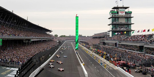
The Penske Entertainment editorial staff is looking back at the 10 biggest moments of 2025 at Indianapolis Motor Speedway in this year-end series, with one installment appearing on the site per day in countdown fashion from Dec. 22-31.