Leinster: J Osborne; T O’Brien, G Ringrose; R Henshaw, J Lowe; S Prendergast, J Gibson-Park; P McCarthy, R Kelleher, T Furlong; RG Snyman, J Ryan; A Soroka, J van der Flier, J Conan (capt).
Replacements: D Sheehan, A Porter, T Clarkson, B Deeny, M…
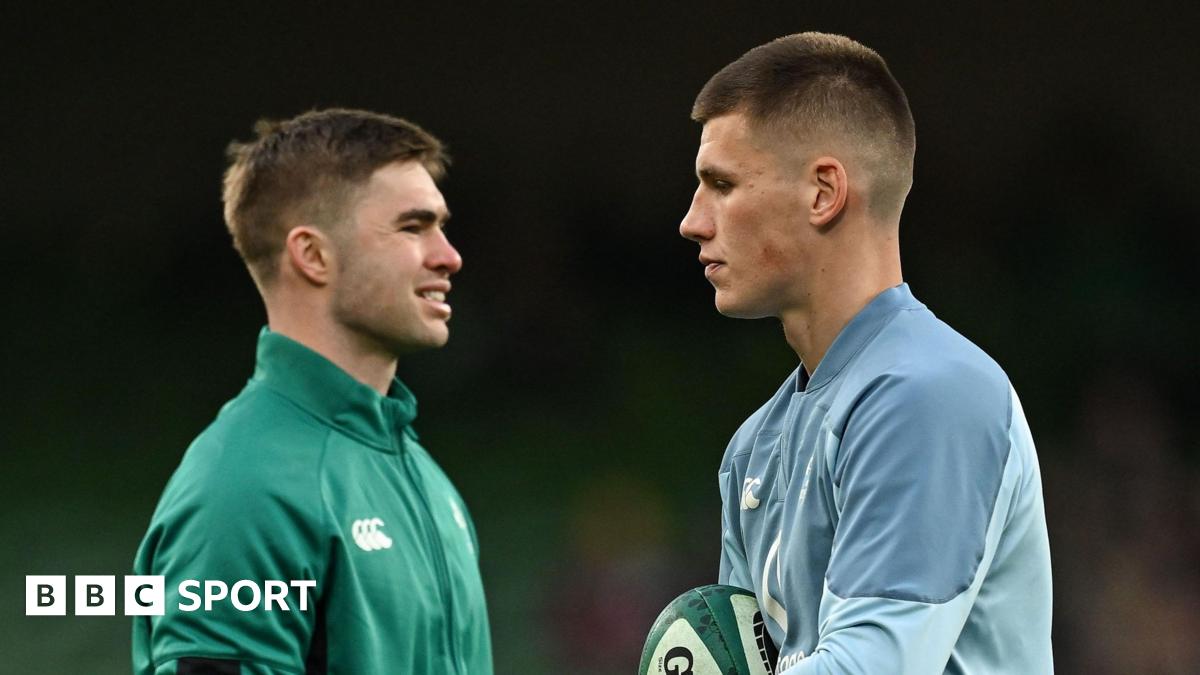
Leinster: J Osborne; T O’Brien, G Ringrose; R Henshaw, J Lowe; S Prendergast, J Gibson-Park; P McCarthy, R Kelleher, T Furlong; RG Snyman, J Ryan; A Soroka, J van der Flier, J Conan (capt).
Replacements: D Sheehan, A Porter, T Clarkson, B Deeny, M…