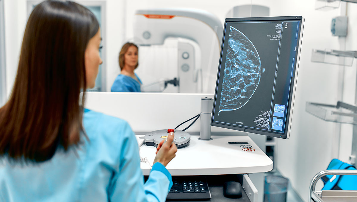
The deep-learning model used the whole “breast architecture,” not just calcification, to predict MACE risk in middle-aged women.
It may one day be possible, through a deep-learning algorithm, to leverage routine mammograms as a way to predict women’s cardiovascular risk, according to retrospective study of Australian data.
The artificial intelligence (AI)-informed tool, like others before it, measures breast arterial calcification (BAC), but it also analyzes other features like breast density and considers age-predicted cardiovascular risk, researchers report in Heart. They say these initial results suggest it works as well as traditional risk equations.
What stands out about the current model, said Jennifer Yvonne Barraclough, PhD (University of New South Wales, Sydney, Australia), is that it’s “the first to use a range of features from mammographic images rather than BAC alone, combined simply with age to predict cardiovascular events.”
BAC is limited in its ability predict such events, Barraclough explained, so their study uses the entire “breast architecture,” with the hope that it’s more reliable. For example, BAC has been shown to be associated with risk of cardiovascular events and certain risk factors, but it is not associated with obesity and inversely correlated with smoking, explain the researchers. The current paper, however, doesn’t directly compare the BAC versus more holistic models, Barraclough added.
Still, the results point to the “potential for a ‘two-for-one’ screening test that does not require any additional data collection beyond screening mammography,” she told TCTMD via email. Age, which informed the model, already is collected at the time of mammography, so this approach would utilize existing resources to ascertain both CV risk and breast cancer.
Women identified as having high CV risk “would then be encouraged to have their CV risk factors assessed and managed accordingly,” said Barraclough.
The question is how such an approach could be implemented in real-world practice, commented Ana Barac, MD, PhD (Inova Schar Cancer and Inova Schar Heart and Vascular, Falls Church, VA).
While a growing body of evidence suggests a connection between BAC and CVD risk, what’s driving the relationship isn’t clear—for example, BAC also doesn’t correlate with coronary artery calcium, she noted.
For this particular model looking at all elements of the mammogram, not just BAC, it makes it hard to understand what the model’s results show about CVD. Barac said it begs the “question of, what are the potential causes? What do we do about that risk once identified?”
She cautioned that women who learn they have high CV risk won’t feel empowered if there aren’t specific steps for prevention, and those told they have low risk might see it as a free pass to not pay attention to their CV health. Despite these caveats, Barac agreed the model predicts CV events with “quite high accuracy.”
The ability to piggyback off of an existing screening tool is also appealing, said Barac. “We are spending a lot of time and effort and sometimes funds in trying to understand individual risk. And this risk might be just already in the mammogram, so why not use it. I think that’s intriguing and positive thinking.”
Model Predicts MACE
For the study, researchers turned to the Lifepool cohort, which enrolled women who had at least one screening mammogram from 2009 to 2020 and agreed to link this screening to health data routinely collected by the Victorian Admitted Episodes Database for hospital admissions and the National Death Index. They worked to develop a deep-learning model based on DeepSurv that uses mammography images to predict extended MACE, defined as either death or a hospital stay for either atherosclerotic cardiovascular disease or heart failure.
Mammograms of 49,196 women (mean age 59.6 years) were used, along with age and radiomic data, to create the model. Over a median follow-up of 8.8 years, 3,392 experienced a first major cardiovascular event, amounting to 7.6 per 1,000 person-years. These events included 2,383 instances of coronary artery disease, 656 MIs, 434 strokes, and 731 cases of heart failure.
The model was able to predict extended MACE with a concordance index of 0.72 (95% CI 0.71-0.73). It performed similarly irrespective of body mass index and across menopausal groups. It offered “similar performance to modern models containing age and clinical variables including the New Zealand ‘PREDICT’ tool and the American Heart Association ‘PREVENT’ equations,” the investigators note.
Barraclough acknowledged that before implementing the model more widely, it needs to be further assessed in other cohorts. There’s also the concern that using mammograms to screen for CV risk might impact existing programs tailored to breast cancer screening.
“We are currently undertaking qualitative research to understand the barriers and enablers for implementation in Australia. Furthermore, this may look very different across different healthcare systems,” she said.
And finally, which specific elements of the breast architecture—such as breast density and microcalcifications—are most closely tied to CV risk haven’t been pinpointed, added Barraclough.
In an editorial, Gemma A. Figtree, MBBS, DPhil, and Stuart M. Grieve, MBBS, DPhil (both from University of Syndney, Australia), call for research to tease out which types of MACE—heart failure, stroke, or atherosclerotic cardiovascular disease—are best predicted by mammogram findings.
“There is uncertainty about the potential mechanism or mechanisms that are reflected by the machine-learning model,” they write. “Does this reflect vascular health and systemic susceptibility to atherosclerosis, or different hormonal or metabolic profiles of the individual?”
Without this understanding, the editorialists caution, it may be difficult to determine the best next steps for women identified as high risk—whether to treat traditional risk factors or, perhaps, refer them for CT angiography. For inspiration, they suggest a potential clinical pathway.
“The application of a new risk tool to triage individuals for screening for subclinical CAD is particularly relevant with the increasing emphasis on atherosclerotic CAD as the disease itself, and heart attacks as more of a catastrophic endpoint. Prospective implementation studies can then be assessed for their ability to reduce the number needed to scan to detect clinically actionable CAD,” Figtree and Grieve conclude.
