Comparison with literature
The analysis of the phenotypes of the pediatric population examined revealed a series of evidence confirming the literature. 90.9% of patients have epistaxis, that is confirmed to be the first symptom to manifest itself during childhood, which, however, rarely requires cauterization or other significant interventions to control bleeding [9], as demonstrated by the absence of major bleedings (0%) in the target population.
36.4% of pediatric patients have mucocutaneous telangiectasias, which, when compared to the prevalence of those in the adult population (100%), has their later presentation confirmed [9]. The same applies to AVMs, present in 27.3% of pediatric patients and 55.6% of adult patients.
The correlation of the phenotype characterized by the presence of pulmonary and cerebral AVMs with genotypes characterized by ENG and ACVRL1 variants is reported in Table 3. The increased frequency of pulmonary and cerebral AVMs in patients – both pediatric and adult – with ENG variants has been confirmed, as reported in literature [8]. However, as evidenced by the presence of cerebral AVMs in 1 pediatric patient (12.5%) with a ACVRL1 variant, it must be reiterated that the genotype alone cannot and must not guide the screening practices, as different AVMs are potentially present in all genotypes [8]. An important pediatric-specific consideration is the higher prevalence of cortical malformations in HHT children, with polymicrogyria described as the most common type [14,15,16].
Analysis of laboratory values
Hemoglobin, serum iron, and ferritin below the respective cutoffs – as referred for age specific pediatric cut-off [11] – were not recorded for any pediatric patient. Moreover, none of these patients needed iron infusions or blood transfusions. These data strengthen published recommendations about blood test timeline both in adults and children with HHT [17]. An apparently normal value of hemoglobin in patients with pulmonary phenotype, as known, may depend on compensatory polycythemia. These patients, therefore, should be focused during the pubertal development period, due to the increase of iron requirement or increased loss – in post-menarche females.
Genotype-phenotype correlation
There is a well-known correlation between genotype and phenotype in HHT. ENG mutations cause severe pulmonary AVMs and a higher incidence of gastrointestinal bleeding, ACVRL1 mutations correlate to milder phenotype with fewer pulmonary complications but significant epistaxis and gastrointestinal issues, while SMAD4 mutations are associated with additional complications, including gastrointestinal polyps and an increased risk of colorectal cancer.
Comparing children’s phenotypes to their parents sharing the same genotype (Table 3), despite the intrinsic limits linked to specific considerations relative to a dynamic pathology, we have seen that, out of a total of 9 families analyzed, a similar clinical phenotype was recorded in 3 families (33.3%).
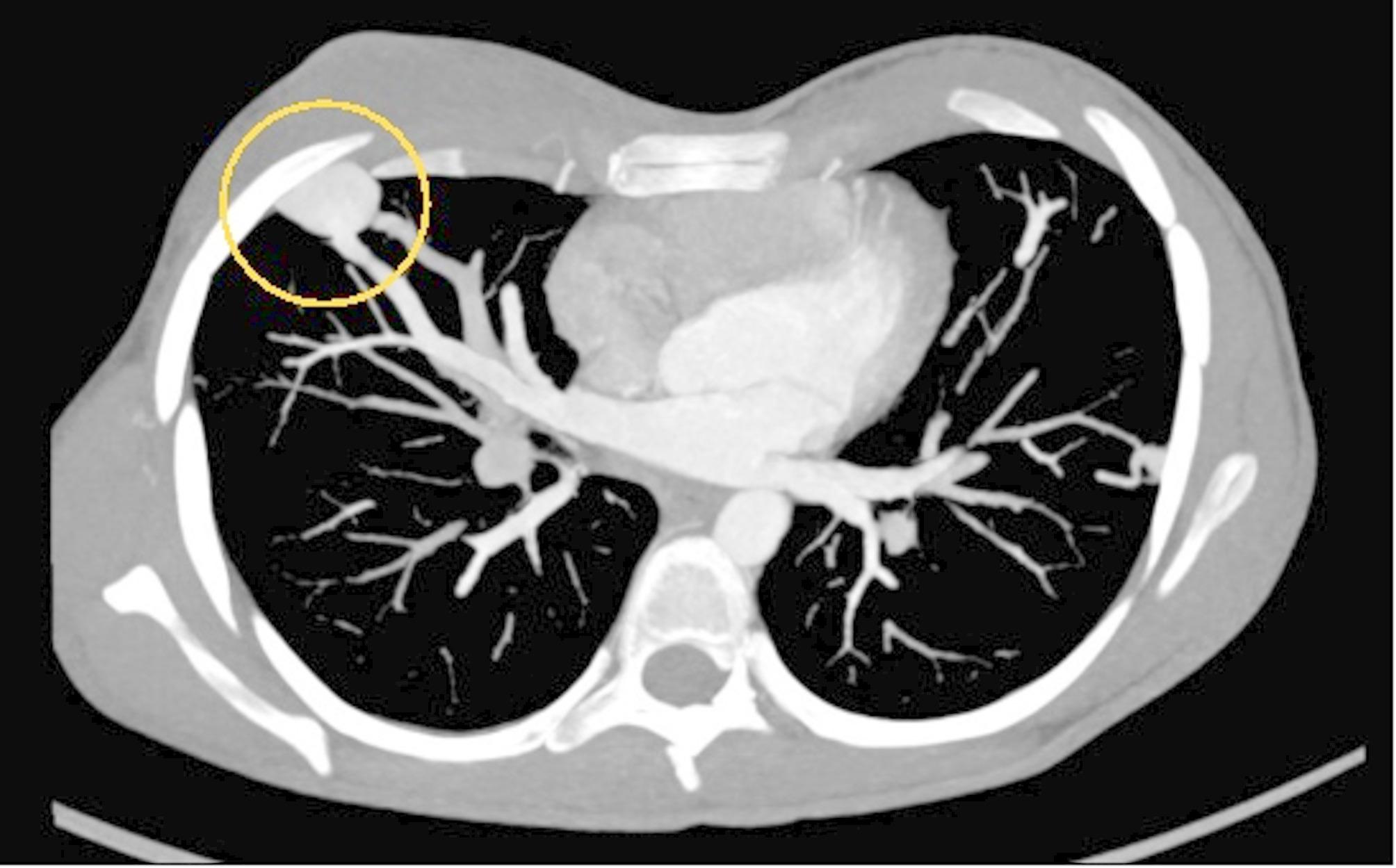
In this regard, the case of family 4 deserves to be reported. The history of this family began in 2018 with the suspicion of HHT in the eldest son following the finding of hypoxemia (SpO2 85%) and multiple pulmonary AVMs, 2 of which were treated with embolization. Further diagnostic investigations were simultaneously carried out in the father and in the second child, and both were found to be affected by HHT with c.230G > A variant in ACVRL1 (shared by all three family members). It is interesting to note that, with the same clinical score (3) assigned through the Curaçao criteria, we were faced with extremely different clinical pictures. The firstborn (20 years) presented epistaxis and the so called “lung phenotype”. The second son (16 years old) also presented epistaxis, but more severe, and, in addition to pulmonary AVMs, a hepatic AVM and a cerebral AVM – i.e. a mixed phenotype. On the other hand, the father (58 years) presented only epistaxis – less severe than in the children – and meager telangiectasias. Therefore, the difference in the phenotypic expression of the disease is evident, as, while it has no impact, if not small, on the quality of life of the father, it has a great impact on that of both children. The story of this family can help reminding that intra-familial phenotypic variation is well-established; in fact, most disease-causing pathogenetic variants in ACVRL1, ENG, and SMAD4 are null alleles that result in haploinsufficiency [18]. Clinicians dealing with families with HHT should keep this peculiarity in mind and eventually discuss it with the families. Clinical variability is expected in the setting of autosomal dominant diseases with variable expressivity, such as HHT. Nonetheless, genotype-phenotype correlations have been well described in adults with HHT [19], but much less in children [20, 21]. In addition, a direct comparison between the phenotypes of parents and children carrying the same gene mutation within the same family has never been performed. This is the added value of our study.
It is important to note that, in Italy, there is a National Health Care System providing a free screening and follow up program for rare diseases, including HHT. This program includes periodic abdominal ultrasound, that can be performed before the symptoms and/or signs of complications might appear.
Our study has some limitations that need to be addressed. This is a single center study including few patients, with observational aims. It follows that, when possible, statistical analysis has been performed but limited by the sample size. A further limitation of our study is the shorter observation period in children compared to their parents. However, it is important to underline that, in recent years, the therapeutic approach for these patients has evolved towards a more conservative strategy, which may partially explain the lower number of treatments observed in children.
Further studies are needed to better investigate these findings.
