Listeria ivanovii subsp. ivanovii is an intracellular bacterium widely distributed in nature, responsible for listeriosis in ruminants and humans [4]. There is no data on the presence of this bacterium in pet animals, to our knowledge, this is the first report on the isolation of L. ivanovii from a cat. In general, there are fewer studies describing the pathogenicity and occurrence of L. ivanovii in animals and the environment compared to L. monocytogenes.
Studies conducted by Cao et al. [13] revealed the presence of L. ivanovii subsp. ivanovii and L. ivanovii subsp. londoniensis in 26 (3.7%) out of 702 fecal samples collected from 25 different species of wild rodents. The authors suggested that the wild rodents could act as the long-term host for L. ivanovii. This is in line with the study of Wang et al., who identified L. ivanovii in seven (20.6%) out of 341 intestinal fecal samples of rodent origin [12]. In addition to L. ivanovii, it is worth mentioning that both studies confirmed the presence of other Listeria species – namely: L. monocytogenes, L. innocua, L. fleischmannii, and L. floridensis – in fecal samples collected from rodents. In Belgium, Bauwens et al. [9] detected L. ivanovii in the feces of red ruffed lemur (Lemur variegatus rubra) and Grauer’s gorilla (Gorilla gorilla graueri) kept in Antwerp Zoo. Additionally, Sarangi and Panda [11] reported the presence of L. ivanovii in a fecal sample collected from a leopard (Panthera pardus). In Poland, this bacterium was also isolated from a red fox (Vulpes vulpes) rectal swab [22].
The presence of L. ivanovii was confirmed in cloacal swabs collected from birds, including chickens, pigeons, ducks, and turkeys. Furthermore, this bacterium was found to contaminate frozen chicken breast fillets [15]. The study conducted by Abuhatab et al. [15] highlights that birds are carriers of L. ivanovii; therefore, further studies are needed to confirm whether these animals can serve as a source of L. ivanovii in the environment.
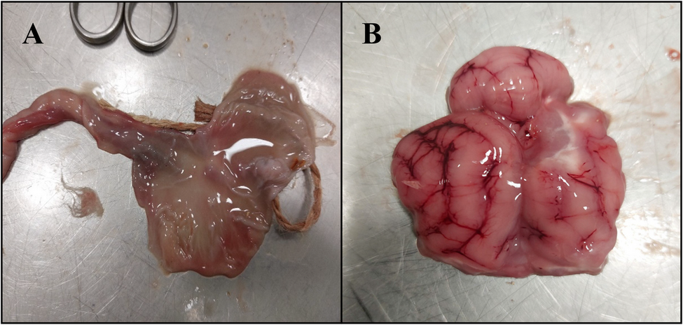
In this study, the source of infection remains unknown. There is no information on the method of feeding and the type of food that was given to the kittens in a litter. It is known that other kittens within this litter died, but they were not diagnosed. Thus, it is not clear why L. ivanovii was present in the cat’s internal organs, but if this bacterium contaminates poultry meat, then such raw food may be a potential source of infection for carnivores, including cats. Additionally, shedding of Listeria in the rodent feces could contaminate food products or food-processing environments by direct or indirect transmission paths.
L. ivanovii was detected in the river and farm water. This suggests that contaminated water may also pose a risk of transmission of this bacterium to animals [14].
Palacios-Gorba et al. [23] identified wild animals, such as deer and wild boars, as potential reservoirs of L. ivanovii, which were detected in their tonsil samples. Moreover, it was shown that L. ivanovii was present in cattle, mainly in the udders. Single isolates were found in tonsils and feces [14, 16]. Furthermore, other studies indicate the presence of L. ivanovii in mastitic milk samples obtained from cattle, buffalo, and goats, suggesting this bacterium’s involvement in udder infections [1, 10].
As was mentioned before, there are only sparse data on the pathogenicity of L. ivanovii, however, unlike L. monocytogenes, which commonly induces meningoencephalitis, L. ivanovii is known not to cause such manifestations in ruminants. Its impact on humans is sporadic, with cases generally restricted to gastroenteritis, bacteremia in immunocompromised individuals, or fetal loss during pregnancy [24]. It should be emphasized that L. ivanovii exhibits a dynamic and complex array of virulence factors, highlighting its evolutionary adaptability [1]. Prominent among these is sphingomyelinase C (SmcL), encoded by the Listeria Pathogenicity Island-2 (LIPI-2), which disrupts host cell membranes and facilitates tissue invasion and immune evasion [25]. Ivanolysin O, a pore-forming hemolysin analogous to listeriolysin O in L. monocytogenes, supports escape from the phagolysosome into the cytoplasm, allowing intracellular survival and replication [1]. The phospholipase SmlC mediates a characteristic shovel-shaped “CAMP-like” response with Rhodococcus equi, which is used for phenotypic identification of L. ivanovii. It was suggested that SmlC contributes to host tropism as it effectively lyses sheep but not horse erythrocytes, which contain less sphingomyelin [1, 26]. Intriguingly, the regulation of virulence factors in L. ivanovii differs from that in L. monocytogenes. Both species rely on PrfA as a central transcriptional regulator, yet L. ivanovii demonstrates strain-specific variations in how these factors are expressed. These differences allow L. ivanovii to adapt its pathogenic potential to specific environmental and host contexts, enhancing its survival and ability to cause a disease [4].
Although listeriosis is rarely reported in cats, a few documented cases of L. monocytogenes infection, involving internal organs, lymphatic and serosal structures, suggest that felines are not resistant to infections by members of the Listeria genus [27, 28]. Therefore, our report confirms the susceptibility of cats to these bacteria and may represent an underrecognized route of L. ivanovii transmission in companion animals. This also suggests that these bacteria can cross species barriers and infect a broader range of hosts, regardless of their usual host specificity.
To our knowledge, no studies described infections caused by L. ivanovii in companion animals, including cats. Nevertheless, this bacterium can cause serious infections and can be isolated from internal organs, as confirmed by Kimpe et al. [29], who identified this microorganism in nodulated liver tissue from a septicaemic chinchilla (Chinchilla lanigera).