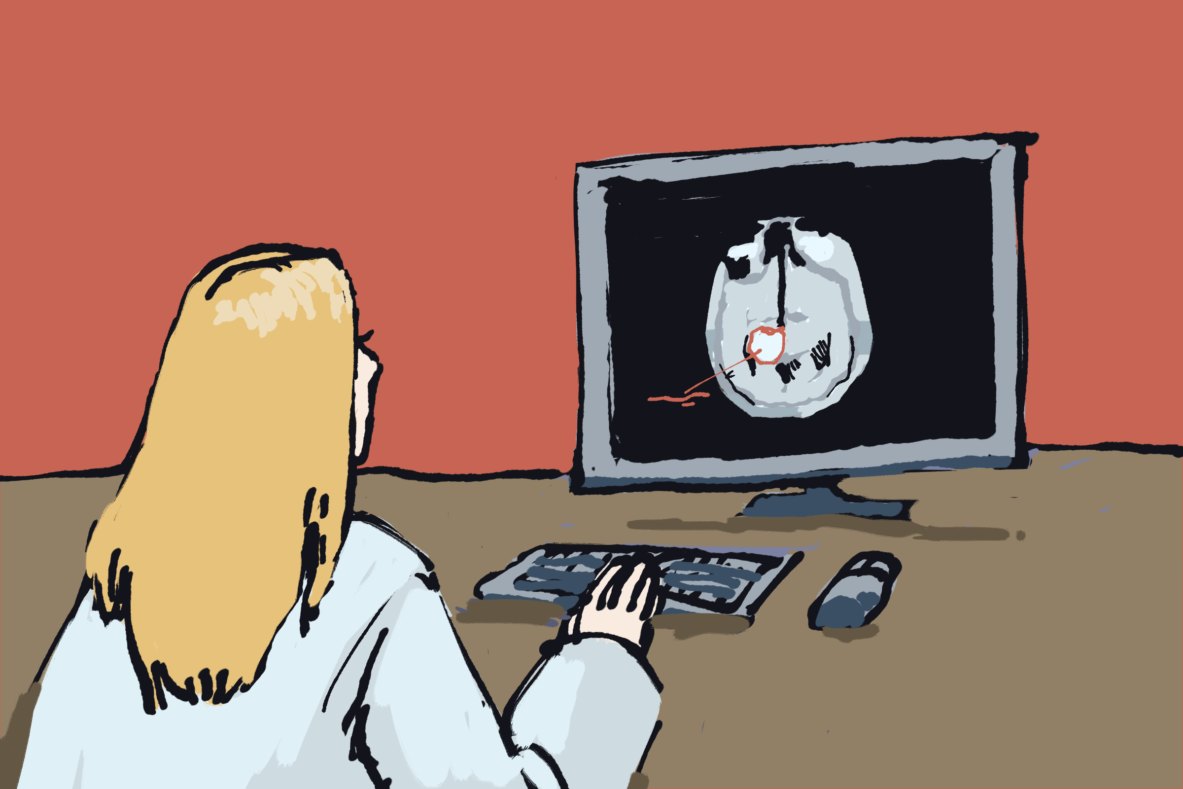
University of Michigan researchers published a study in September, revealing that brain tumors rewire the use of glucose to fuel growth. By removing a specific amino acid from a patient’s diet, tumor growth slows down, making alternative treatments more effective. Published in the science journal Nature, the paper focused on the ways glioblastomas, the most deadly form of malignant brain tumor, use glucose differently than a healthy brain does.
In an interview with The Michigan Daily, Wajd Al-Holou, Michigan Medicine neurosurgeon, assistant professor and co-author of the research, said the study arose from questions surrounding the tumors’ ideal environment and how to dismantle the elements within that environment. The study utilized both mouse models and eight patients undergoing surgery for suspected glioblastomas. Al-Holou emphasized the importance of the study moving from mouse models to human brains.
“In order to develop new treatments, we really have to study how these tumors behave in patients and try to find ways to attack the tumors,” Al-Holou said. “One of the ways that we have been working on is trying to understand the nutritional needs of the tumors. If we can understand what tumors need to grow, then we can essentially block that nutrient from getting in.”
Healthy brain tissue uses glucose to make an amino acid serine to function properly. The study shows brain tumors thrive on serine, but the cancer cells within them are unable to produce serine like healthy brain tissue. As a result, those cancer cells can only use the blood’s pre-existing serine.
In an interview with The Daily, Daniel Wahl, associate professor of radiation oncology and co-author of the research, said the team removed serine from the circulatory system of mice with tumors and applied that data to human patients. He found that brain tissue was still able to produce the serine it needed, but the brain tumors thrived off of the excess serine in the bloodstream. The researchers also found that when serine was removed from a group of mice’s diet, tumors grew at a slower rate and became more sensitive to existing brain tumor treatments.
“We put them on diets that didn’t have any of this amino acid serine,” Wahl said. “It had protein, just no serine, and that slowed down the tumor growth in the mice, in many of the mouse models we used, and made our normal treatments like radiation and chemotherapy work better.”
Glioblastomas are typically treated with radiation therapy and chemotherapy, but the tumors usually return within nine to 12 months and the survival rate is low. In the study, researchers applied the findings from the mouse models to human patients to identify the effect of serine removal in humans. If companies can manufacture diets for humans that exclude serine, these diets may be able to slow tumor growth and make radiation therapies and chemotherapy significantly more effective, according to the study.
This study marks a significant step forward for brain tumor research, given that the last major drug advancement in the treatment of glioblastomas that used temozolomide came out in 2005. Al-Holou said it is challenging to remove serine from food, but researchers are working on methods to begin clinical trials as soon as possible.
“Unfortunately, (serine is) essentially found in every protein source out there,” Al-Holou said. “The next steps involve developing a diet where we can remove serine. That’s a complicated process, but something we’re working on, and we’ve partnered with a company to make it happen.”
The current study focuses on glioblastomas, a specific form of brain tumor, but Wahl says his team are already studying other types of brain tumors to expand the scope of their research. Of the eight human patients in their study, two turned out to have another form of brain tumor that was not than glioblastomas. This difference has led to research into how the serine-free diet could be applied to other kinds of brain cancer.
“Now we are purposefully studying other types of brain cancer with this technique — some slower-growing tumors, some tumors with other types of molecular genetics, some pediatric brain cancers,” Wahl said. “So we really think that this is a technique that (if) we have worked it out, we can gain a lot of new information about different tumor types.”
In an interview with The Daily, Rackham student Baharan Meghdadi, co-author of the study , said the main task of the engineering branch of the team was to create statistical models that could make predictions about how glioblastoma cells rewire nutrient pathways and how these changes could impact response to medical treatments. With these models, the nutritional needs of tumors and the paths the tumor uses to obtain these nutrients can be compared to those of healthy cells.
“(This data) comes from the experiment, but to see the differences in what’s going on in the tumor cells and the healthy cells, it’s good to have some models that can recapitulate the metabolism in these cells,” Meghdadi said. “It can also help us to make some hypotheses and then test them experimentally. These models include the different routes that glucose can be used in cells.”
Al-Holou said the researchers were only able to come to these useful conclusions because of the cross-departmental nature of their work. Collaborators worked on the study from across Michigan Medicine, the College of Engineering and other universities and companies.
“The lack of major advancements in brain tumor research has been because a lot of people are kind of working in silos on their own,” Al-Holou said. “What we’ve done here is combine efforts from radiation oncology, neurosurgery, metabolomics and engineering together to come up with this. It could not have been done with one person, or with one lab, or one group alone.”
Daily Staff Reporter Grace Schuur can be reached at gschuur@umich.edu.