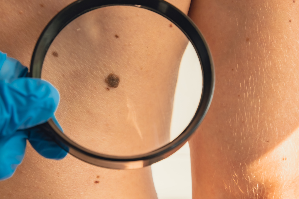
Performance of a convolutional neural network in determining differentiation levels of cutaneous squamous cell carcinomas was on par with that of experienced dermatologists, according to the results of a recent study published in JAAD International.
“This type of cancer, which is a result of mutations of the most common cell type in the top layer of the skin, is strongly linked to accumulated [ultraviolet] radiation over time. It develops in sun-exposed areas, often on skin already showing signs of sun damage, with rough scaly patches, uneven pigmentation, and decreased elasticity,” stated lead researcher Sam Polesie, MD, PhD, Associate Professor of Dermatology and Venereology at the University of Gothenburg and Practicing Dermatologist at Sahlgrenska University Hospital, both in Gothenburg, Sweden.
Background and Study Methods
Preoperative punch biopsies are not typically performed when a patient has a suspected squamous cell carcinoma; the specimen is usually sent for histopathological analysis after being excised.
Researchers trained a de novo convolutional neural network to discriminate between well, moderately, and poorly differentiated cutaneous squamous cell carcinomas. The model was trained on 1,829 clinical close-up images of cutaneous squamous cell carcinoma, including 68.6% of well-differentiated cases, that were divided into training (n = 1,329), validation (n = 200), and test sets (n = 300). Then, the model’s performance was compared to the combined assessment of seven independent dermatologists’ reads. The dermatologists also specified how certain they were about their estimated level of differentiation for each case and noted which clinical features were present in each tumor.
Key Study Findings
The convolutional neural network model had an area under the curve of 0.69 (95% confidence interval [CI] = 0.63‒0.76) and the dermatologists’ combined assessment had an area under the curve of 0.70 (95% CI = 0.64‒0.76; P = .79), with moderate agreement between their reads, which highlights the complexity of the task.
Ulceration (odds ratio [OR] = 2.34; 95% CI = 1.16‒4.72) and flat surface topography (OR = 2.94; 95% CI = 1.23‒7.01) were more common in moderately or poorly differentiated tumors.
“We believe the use-case presented here is a promising machine-learning approach worth pursuing, as it has the potential to assist and augment dermatologists in preoperative decision-making—helping to determine appropriate surgical margins or assess whether alternative, less invasive treatments might be suitable,” the study authors concluded.
The model needs to be refined and evaluated further, though, before it can be effectively used to assist dermatologists in clinical practice, investigators noted.
Disclosure: For full disclosures of the study authors, visit sciencedirect.com.
