A recent conference celebrated the creation of the first prototype of the spectral optical coherence tomography (SOCT) instrument. Introduced in 2000, the prototype, which was developed at the Institute of Physics of Nicolaus Copernicus University in Toruń, Poland, was the basis for the world’s first commercial SOCT device introduced in 2004, according to a press release issued by the OFTALMIKA Eye Clinic, Bydgoszcz, Poland.
To celebrate this landmark innovation in ocular diagnostics, the OFTALMIKE Eye Clinic hosted a conference titled, “OCT in Ophthalmology: From Science to Practice” on September 26 to 27, 2025, in Toruń. The conference highlights included discussions of the advances in diagnosing and treating geographic atrophy, new therapies for macular diseases, wide-angle imaging for retinal disorders, OCT in vitreoretinal interface assessment, and OCT in glaucoma diagnostics, the press release enumerated.
Professor Andrzej Kowalczyk and Professor Maciej Wojtkowski were listed as the guest speakers at the conference, where they received an award in recognition of their contributions to ophthalmic diagnostics.
The prototype SOCT machine paved the way for the subsequent iterations of the technology. When the technology entered the commercial market in 2004, via collaboration with the late Prof. Józef Kałużny, MD, PhD, it “quickly entered clinical practice, extending ophthalmic diagnostics to corneal imaging and biomechanics,” the press release stated.
Rapid technologic advancements
Following the assembly of the prototype, the OCT researchers were able to present the first high-resolution images of the eye of a healthy volunteer just 2 years later, Professor Jakub Kałużny, MD, PhD, recounted. He is Medical Director of the OFTALMIKA Eye Hospital.
Underscoring the early research efforts into the new technology, the OCT devices have become indispensable in clinics and research centers across all continents, while Toruń scientists continue to pioneer portable and interdisciplinary applications of this technology, the press release related.
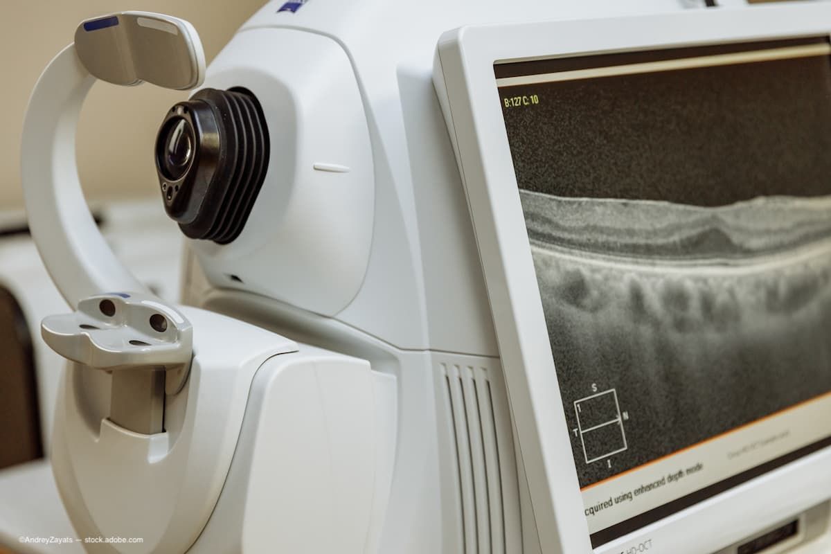
“Often compared to ‘optical histology,’ OCT provides highly detailed images of the retina, cornea, and optic nerve. It enables early detection and monitoring of glaucoma, age-related macular degeneration, diabetic retinopathy, and many other eye conditions,” the press release related. Adding to that, Professor Jakub Kaluzny emphasized, “OCT has become the global gold standard in ophthalmology diagnostics, transforming how we care for vision health.”
Prof. Bartłomiej Kałużny, MD, PhD, also pointed out the importance of the instrument to the anterior segment, “The technology also plays a vital role in imaging the anterior segment of the eye. “With OCT, we can quantify corneal and lens pathologies and evaluate the drainage angle — crucial for surgical qualification and treatment monitoring.”