While corneal tattooing is one of the most widely performed cosmetic procedures and is also used for therapeutic purposes, it is not without complications. The literature describes adverse effects of infectious keratitis, granulomatous inflammatory inflammatory reactions, and pigmentary alterations such as fading, discoloration, and unintended migration. These complications could stem from immune responses, instability of the pigment, or the technique employed during the procedure [4]. While most reported cases focus on ocular complications, cutaneous pigmentary changes following corneal tattooing remain undocumented, making our case a unique contribution to the existing literature.
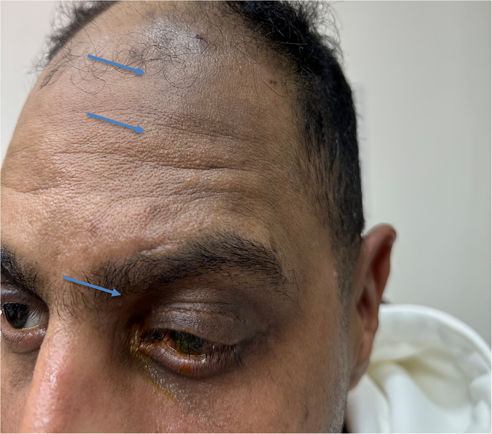
Although the tattoo pigment was originally limited to the corneal, we believe that the pigment may have migrated through lymphatic channels or along the perineural pathways of the ophthalmic (V1) branch of the trigeminal nerve. The pattern of periocular skin pigmentation closely followed the V1 dermatome, supporting the possibility of perineural spread or transport via perivascular lymphatics, particularly after local tissue disruption from the augmentation procedure. In a patient with naturally darker skin, such pigmentation may have been less obvious at first, becoming more noticeable as it extended beyond the immediate periorbital region. While this remains a proposed explanation, similar patterns of pigment migration have been described in cases following periocular tattooing.
Lee et al. [5] reported cases of pigment fanning after eyelash and eyebrow tattooing, where pigment migrated beyond the intended area, leading to infraorbital and periorbital pigmentation. They hypothesized that this phenomenon occurs due to deep pigment placement in the skin, allowing pigment to spread through the lymphatic system or subcutaneous tissue planes. Additionally, variations in skin thickness, gravitational effects, and localized inflammatory responses were considered contributing factors.
This in line with our case, where corneal tattooing resulted in unexpected pigment migration, manifesting as cutaneous hyperpigmentation following the distribution of the ophthalmic (V1) division of the trigeminal nerve. In contrast to the cases described by Lee et al., where pigment remained within the periocular region, our case suggests a more extensive pattern of pigment dispersion, possibly involving neural pathways or deeper lymphatic drainage routes [6]. Given the patient’s history of multiple ocular surgeries and prior orbital trauma, it is plausible that disrupted anatomical barriers, altered tissue integrity, or changes in vascular dynamics played a role in facilitating the pigment’s unexpected migration.
Regarding the stability of tattoo color, Calas et al. [7] investigated the long-term color stability of corneal tattoos, reporting three cases where the tattoo color significantly changed between 5 and 6 years postoperatively, shifting from black to golden-brown. Through a retrospective analysis, they identified the presence of iron-based pigments, which were likely oxidized into golden-brown ferric iron oxide due to oxygen exposure in the aqueous environment. The study also noted that corneal edema in some cases may have contributed to the oxidation process. Despite these pigmentary changes, corneal tattooing remained a valuable technique for reducing photophobia in patients with aniridia or corneal opacities.
Sharma et al. [8] reported a case of granulomatous keratitis following corneal tattooing, an uncommon complication that supports the hypothesis that immune reactions may contribute to pigment-related complications, such as trigeminal nerve-distributed hyperpigmentation in our patient.
In clinical practice, various techniques are used to introduce pigment into the corneal stroma, depending on the patient’s ocular condition and the surgeon’s preference. The most commonly used technique is intrastromal injection, where pigment is introduced directly into the corneal stroma using a fine needle under microscope or slit-lamp guidance. This method is popular because it offers deep and long-lasting color, especially in eyes with dense corneal opacities. However, it does require precision—if the injection is too superficial or too deep, there’s a chance that the pigment might migrate unevenly or spread beyond the intended area, particularly in patients with prior surgeries or compromised corneal tissue [9].
The lamellar pocket technique offers a more refined approach, especially when dealing with deeper corneal scars. In this method, a mid-stromal pocket is carefully created using a crescent blade or femtosecond laser, and the pigment is placed into this confined space. This technique helps trap the pigment securely and reduces the likelihood of it leaking or migrating, making it ideal for patients who need stable, long-term results [10].
Femtosecond-assisted tattooing is a more modern, high-precision approach that allows surgeons to create perfectly shaped intrastromal channels at consistent depths. Because of its accuracy, it’s associated with excellent pigment containment and very low risk of unintended pigment distribution [10].
In our case, we used the intrastromal injection technique. However, previous studies do not specify the exact method used to apply the pigment to the cornea or how it may have contributed to pigment spread. We believe that the technique itself could play a role in how the pigment behaves, and that different methods may influence the risk of pigment disruption or migration.
Various inks are used in corneal tattooing, each with different risks. India ink is the most common—it’s stable but can fade or migrate if not placed properly [3]. Rotring ink is low-cost but not designed for the eye, raising safety concerns [9]. Metallic pigments like gold and silver are outdated due to poor results and toxicity [2]. Iron oxide inks may change color over time [7]. Although rare in corneal tattooing, pigment migration has been seen more often in cosmetic periocular tattoos, where pigment can spread if placed too deeply, especially in areas with thinner or damaged tissue ([5]– [6]).
Despite the of method used or type of ink, disruption of periocular tattoos has been reported. Goldberg et al. [11] and Sadoughi et al. [12] described cases of inadvertent corneal pigmentation following cosmetic eyelid tattooing, where pigment unintentionally entered the corneal stroma.
Recent literature has drawn attention to the risk of pigment migration into the conjunctiva and surrounding tissues during corneal tattooing. Yilmaz and colleagues [13] reported that performing corneal tattooing at the same time as other ocular procedures may increase this risk, especially if the conjunctiva is compromised. They advise against combining such surgeries to reduce the chance of pigment spread and other complications. In our case, the augmentation was performed as a stand-alone procedure, with no concurrent ocular surgeries, which may have helped limit further dirct dissemination of the pigment to the conjunctiva.
Symptoms included ocular discomfort and redness shortly after the procedure. While one case required partial surgical removal, both were managed conservatively with gradual resolution. These reports highlight the risk of pigment penetration during periocular tattooing, emphasizing the need for precise technique to prevent ocular complications.