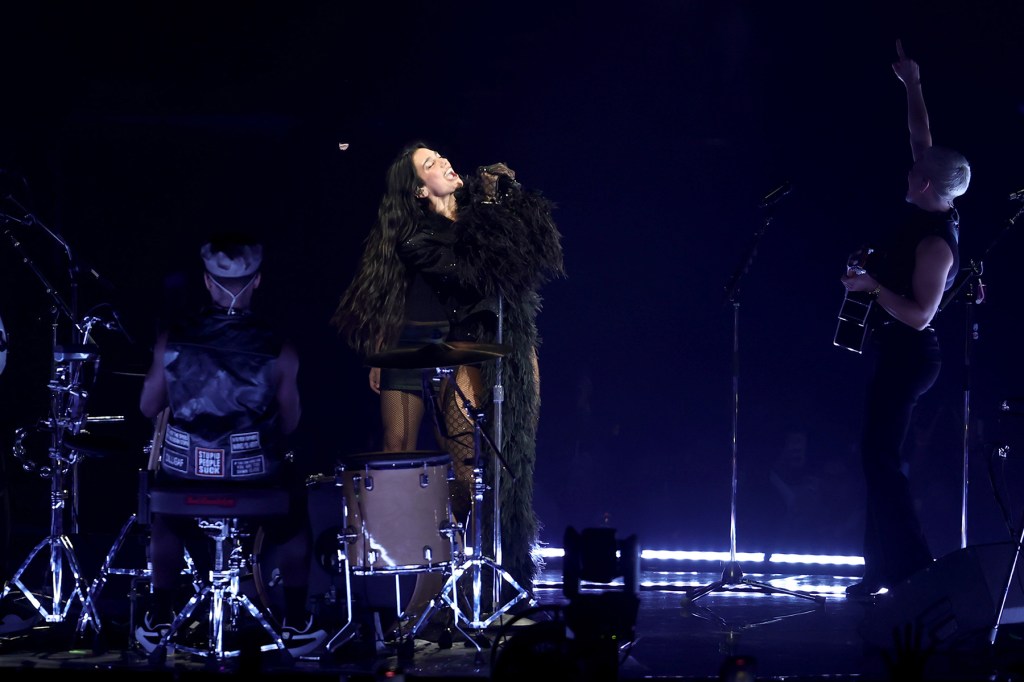
K. pneumoniae-induced septic embolism and prostatic abscesses in a treatment-naive type 2 diabetic patient: a case report | BMC Infectious Diseases

The patient sought medical attention a week ago due to sudden onset of generalized fatigue, dysuria, fever, rectal tenesmus, and constipation. The febrile episodes were characterized by recurrent spikes (39.4 °C) and rigors, notably without accompanying cough, sputum production, diarrhea, or cutaneous eruptions. Based on the provisional diagnosis of “hepatic malignancy with pulmonary metastases and superimposed infection” established at the local hospital, the patient received triple antimicrobial therapy with cefazolin sodium (1.5 g q8h IV) + moxifloxacin (0.4 g qd IV) + ornidazole (0.5 g q12h IV). The patient showed no clinical improvement, with persistent signs of sepsis and hypotension, ultimately necessitating transfer to our tertiary center’s ICU for further management.

On admission, the patient appeared critically ill with tachypnea (respiratory rate 30/min), facial flushing, fever (38.9 °C), blurred mind, hypotension (BP 86/55 mmHg), and a pulse of 106 bpm. His qSOFA score was 3 and Glasgow Coma Scale score was 11 (E3, V4, M4). Pulmonary auscultation identified globally diminished breath sounds accompanied by coarse moist rales throughout all lung fields, particularly pronounced in bilateral lower zones. Abdominal inspection noted significant distension with marked tenderness localized to the right upper quadrant, where hepatic and renal angle percussion elicited reproducible pain; notably absent were peritoneal signs or shifting dullness. Bilateral lower extremities exhibited grade 2 pitting edema extending to mid-calf level. Rectal examination detected a 3 × 4 cm soft, exquisitely tender mass occupying the anterior rectal wall, demonstrating localized fullness without evidence of sphincter compromise. In addition, the patient had a 5-year history of type 2 diabetes mellitus (T2DM) that was completely untreated, with no documented history of glycemic monitoring or pharmacologic intervention. Point-of-care (POC) blood glucose testing showed a concentration of 18.2 mmol/L. The patient is administered 8 units of insulin Neutral Protamine Hagedorn daily at 10 PM and 8 units of insulin aspart before breakfast, lunch, and dinner (30 min prior to each meal). Blood glucose is monitored every 2 h with the goal of maintaining levels within normal limits.

The arterial blood gas showed pH 7.48, FiO₂ 41% with electrolytes Na⁺ 129 mmol/L, K⁺ 4.2 mmol/L, Cl⁻ 103 mmol/L. Complete Blood Count shows critical leukocytosis (white blood cell 30.93 × 10⁹/L) with severe anemia (hemoglobin 89 g/L), neutrophilia (absolute neutrophil count 15.64 × 10⁹/L), and decreased red blood cell count (2.95 × 10¹²/L). Biochemistry: Marked abnormalities include albumin 20.8 g/L, C-reactive protein 154 mg/L, and procalcitonin 5.9 ng/mL, with low total protein (54 g/L), alanine aminotransferas (8.8 U/L), and uric acid (119 µmol/L). Urinalysis shows 2 + protein, 2 + white blood cells, and 4 + glucose in the patient’s urine. The patient received empiric imipenem/cilastatin 500 mg q6h + vancomycin 1 g q12h with enoxaparin 1 mg/kg q12h, Fluid resuscitation and nutritional optimization.

Contrast CT scan Showed clots were seen in the right liver vein (Fig. 1A) and left kidney vein (Fig. 1B). Multiple low-density lesions with rim enhancement in the prostate (Fig. 1C) and right liver (Fig. 1A), likely abscesses. Mildly enlarged lymph nodes noted in both groin areas. There were bilateral patchy shadows and nodules in the lungs, a small amount of pleural effusion in the thoracic cavity (Fig. 1D). The cranial CT scan shows no abnormalities in the patient’s brain. The preliminary diagnosis was sepsis and septic embolism (in the right hepatic/left renal vein) secondary to prostatic and hepatic abscesses. Under ultrasound guidance, percutaneous drainage of the right hepatic lobe and transperineal prostatic drainage were sequentially performed, yielding a significant amount of purulent fluid, with subsequent placement of an indwelling catheter in the right hepatic lobe. The drained fluid was sent for bacterial culture and metagenomic next-generation sequencing (mNGS) analysis for pathogen identification.

Fig. 1

Patient’s CT findings on admission. The patient exhibits hypodense lesions in the right lobe of the liver (A), left kidney (B), and prostate (C). Filling defects are observed in the right hepatic (A) and left renal vein (B). Additionally, there are ground-glass opacities, patchy shadows, and nodular shadows in both lungs (D). Red arrows: Filling defects. Black arrows: hypodense lesions

KP was concordantly detected across blood culture, purulent fluid culture, and mNGS. Furthermore, mNGS analysis detected the presence of resistance genes to third-generation cephalosporins and penicillins in the identified Klebsiella pneumoniae strain. Therapy de-escalated to imipenem monotherapy. Following a two-week targeted therapy regimen, the patient exhibited significant clinical improvement with concomitant normalization of laboratory parameters. Radiological assessment further revealed complete resolution of the septic embolism (Fig. 2A-B). Contrast-enhanced imaging revealed substantial abscess regression (Fig. 2A-C). Concurrent thoracic imaging showed resolving pulmonary infiltrates and minimal residual pleural effusions (Fig. 2D), prompting discharge with scheduled surveillance.

Fig. 2
figure 2

Patient’s CT findings at discharge. The patient’s imaging findings have significantly improved. The hypodense lesions in the liver (A), kidney (B), and prostate (C) have shown notable resolution. Filling has been restored in the right hepatic (A) and left renal vein (B). Furthermore, the lung tissue has returned to a normal appearance (D). Yellow arrows: Venous filling

At the 3-month follow-up after discharge, the patient was satisfied with the results of the treatment and has resumed his normal life. The ultrasound examination indicated that the prostate had returned to normal (Figure S1). After adhering to the doctor’s instructions, the patient’s blood glucose levels have been successfully controlled within the normal range. There were no adverse events throughout the process.

Continue Reading