Cardiac MRI shows the effects of air pollution on the heart — and the findings aren’t good, according to a study published July 1 in Radiology.
The research adds to growing evidence that air pollution is a cardiovascular risk factor, contributing to residual risk not accounted for by typical clinical predictors such as smoking or hypertension, the RSNA noted in a statement.
“Even modest increases in air pollution levels appear to have measurable effects on the heart,” said senior author Kate Hanneman, MD, of the University of Toronto in Canada. “Our study suggests that air quality may play a significant role in changes to heart structure, potentially setting the stage for future cardiovascular disease.”
Heart disease is the main cause of death around the world, the investigators noted. Although prior work has indicated that poor air quality contributes to cardiovascular disease, changes in the heart resulting from exposure to air pollution have remained unclear, they explained, observing that fine particulate matter in the air may contribute to diffuse myocardial fibrosis, a form of scarring in the heart muscle that can precede heart failure. (“Fine particulate matter” can include vehicle exhaust, industrial emissions and wildfire smoke, and particles are small enough to enter the bloodstream through the lungs.)
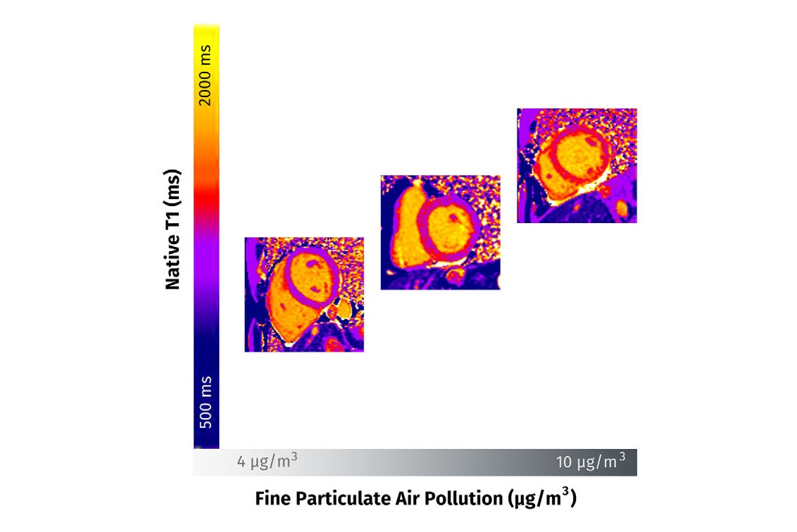
With Hanneman, a team led by Jacques Du Plessis, MD, also of the University of Toronto, explored the relationship between long-term exposure to fine particulate matter with 2.5-µm or smaller aerodynamic diameter (PM2.5) and the extent of diffuse myocardial fibrosis quantified with cardiac MRI native T1 mapping z scores (used to assess myocardial tissue characteristics; score expressed as zero, positive, and negative, with positive scores indicating potential abnormality).
The study included a total of 694 patients who underwent cardiac MRI between January 2018 and December 2022. Of these, 493 had dilated cardiomyopathy and 201 had normal findings. Du Plessis’ team quantified diffuse myocardial fibrosis and assessed patients’ residence-specific ambient PM2.5 concentration (the mean of daily exposure concentration in the year before cardiac MR imaging using measurements from the nearest air quality monitoring station).
The group reported the following:
- In multivariable models, each 1-µg/m3 increase in one-year mean PM2.5 exposure was associated with a 0.3 higher native T1 z score in patients with dilated cardiomyopathy (p < 0.001) and 0.27 higher native T1 z score in controls (p = 0.02).
- For absolute values, each 1-µg/m3 increase in one-year mean PM2.5 exposure was associated with 9.1 msec higher native T1 at 1.5-tesla imaging (p = 0.01) and 12.1 msec higher native T1 at 3-tesla imaging (p < 0.001). (An elevated native T1 typically indicates abnormal changes in the myocardial tissue composition.)
- The largest effect sizes for the association of PM2.5 exposure with native T1 z scores were in women (p < 0.001), smokers (p = 0.04), and patients with hypertension (p = 0.004).
Overall, higher long-term exposure to fine particulate air pollution was connected to higher levels of myocardial fibrosis in both the patients with cardiomyopathy and the controls, suggesting that “myocardial fibrosis may be an underlying mechanism by which air pollution leads to cardiovascular complications,” the authors wrote.
How can clinicians gauge a patient’s cardiac health in relation to poor air quality? Neighborhood data can help, according to Hanneman.
“A simple way to estimate someone’s exposure to air pollution is by looking at air quality data for the neighborhood they live in — information that is publicly available in many areas,” she told AuntMinnie. “Some health providers may ask about environmental exposures during routine visits. For people who want a more detailed assessment, portable air monitors are available that provide real-time air quality data.”
In any case, the effects of air pollution on lung health can be mitigated both on individual and societal levels, Hanneman noted.
“On a personal level, people can take steps like limiting outdoor activity on days when air quality is poor and using indoor air purifiers to reduce exposure,” she said. “On a broader scale, public health efforts include improving air quality standards, reducing emissions from traffic and industry, and addressing sources such as wildfire smoke to improve air quality for everyone.”
In an accompanying commentary, Davis Vigneault, MD, DPhil, of Stanford University emphasized the role radiology can play in tackling the problem of air pollution exposure and heart disease risk.
“The study … suggests several interesting avenues for future research, including the investigation of particulate pollution components, related co-pollutants, and at-risk subgroups,” he noted. “Moreover, [the] work sets an important example of how imaging research may be used beyond the diagnosis of an individual patient to guide public policy interventions to improve health outcomes more broadly.”
The complete study can be found here.