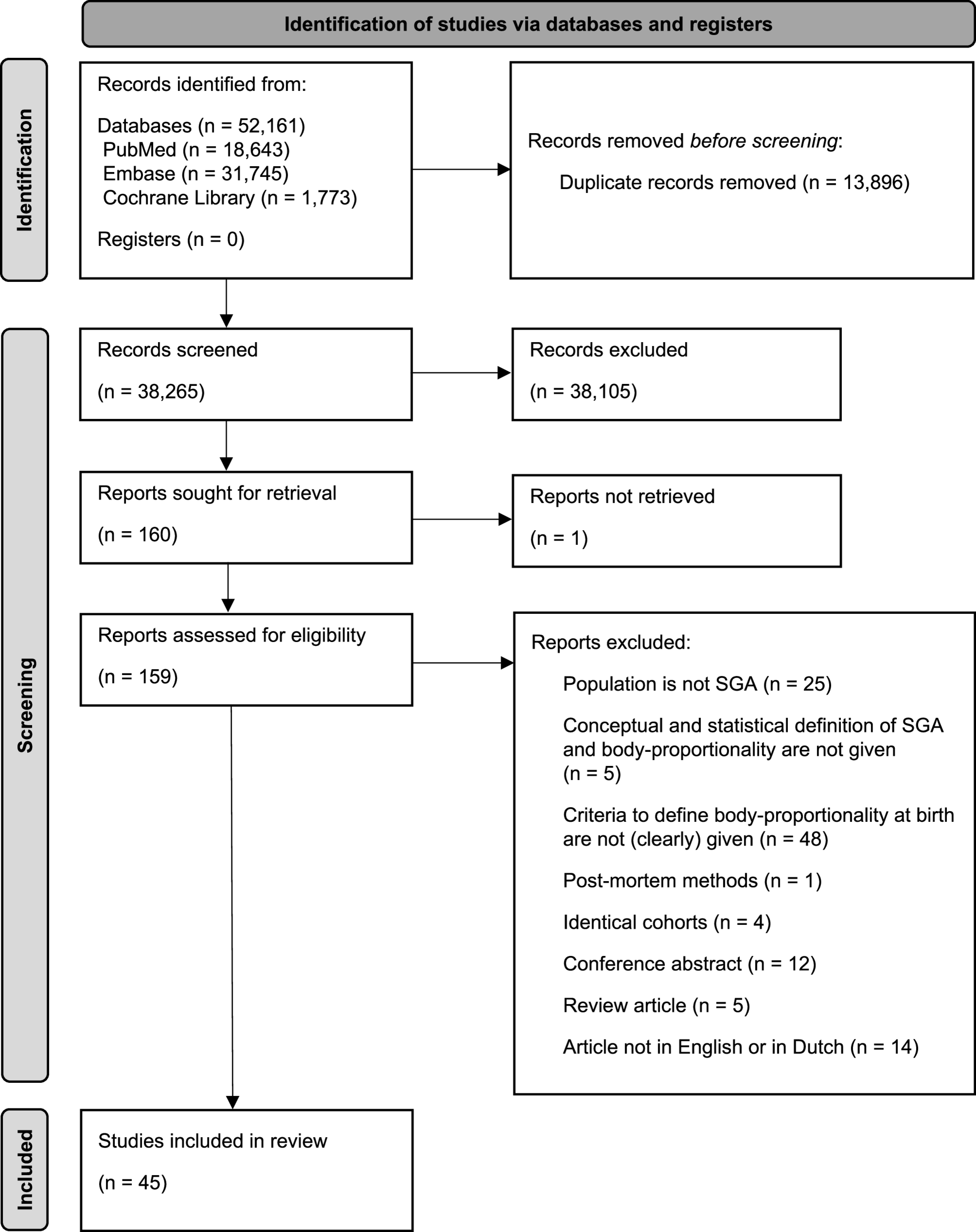
Vayssière C, Sentilhes L, Ego A, Bernard C, Cambourieu D, Flamant C, et al. Fetal growth restriction and intra-uterine growth restriction: guidelines for clinical practice from the French college of gynaecologists and obstetricians. Eur J Obstet Gynecol Reprod Biol. 2015;193:10–8.
Google Scholar
Battaglia FC, Lubchenco LO. A practical classification of newborn infants by weight and gestational age. J Pediatr. 1967;71:159–63.
Google Scholar
Beune IM, Bloomfield FH, Ganzevoort W, Embleton ND, Rozance PJ, van Wassenaer-Leemhuis AG, et al. Consensus based definition of growth restriction in the newborn. J Pediatr. 2018;196:71-6.e1.
Google Scholar
Hughes MM, Black RE, Katz J. 2500-g low birth weight cutoff: history and implications for future research and policy. Matern Child Health J. 2017;21:283–9.
Google Scholar
Landmann E, Reiss I, Misselwitz B, Gortner L. Ponderal index for discrimination between symmetric and asymmetric growth restriction: percentiles for neonates from 30 weeks to 43 weeks of gestation. J Matern Fetal Neonatal Med. 2006;19:157–60.
Google Scholar
Bhat MA, Kumar P, Bhansali A, Majumdar S, Narang A. Hypoglycemia in small for gestational age babies. Indian J Pediatr. 2000;67:423–7.
Google Scholar
Geva R, Eshel R, Leitner Y, Fattal-Valevski A, Harel S. Memory functions of children born with asymmetric intrauterine growth restriction. Brain Res. 2006;1117:186–94.
Google Scholar
Bocca-Tjeertes I, Bos A, Kerstjens J, de Winter A, Reijneveld S. Symmetrical and asymmetrical growth restriction in preterm-born children. Pediatrics. 2014;133: e650–6.
Google Scholar
Chard T, Costeloe K, Leaf A. Evidence of growth retardation in neonates of apparently normal weight. Eur J Obstet Gynecol Reprod Biol. 1992;45:59–62.
Google Scholar
Levit Y, Dym L, Yochpaz S, Manor Y, Adler A, Halutz O, et al. Assessment of risk indicators for targeted cytomegalovirus screening in neonates. Neonatology. 2020;117:750–5.
Google Scholar
Gordijn SJ, Beune IM, Thilaganathan B, Papageorghiou A, Baschat AA, Baker PN, et al. Consensus definition of fetal growth restriction: a Delphi procedure. Ultrasound Obstet Gynecol. 2016;48:333–9.
Google Scholar
Karadavut B, Smits I, Van Dillen J, Hogeveen M. The criteria to classify body-proportionality of the small for gestational age newborn: a scoping review protocol. OSF Preprints. 2021. https://doi.org/10.31219/osf.io/wnjhe.
Peters MDJ, Marnie C, Tricco AC, Pollock D, Munn Z, Alexander L, et al. Updated methodological guidance for the conduct of scoping reviews. JBI Evid Synth. 2020;18:2119–26.
Google Scholar
Tricco AC, Lillie E, Zarin W, O’Brien KK, Colquhoun H, Levac D, et al. PRISMA extension for scoping reviews (PRISMA-ScR): checklist and explanation. Ann Intern Med. 2018;169:467–73.
Google Scholar
McGowan J, Sampson M, Salzwedel DM, Cogo E, Foerster V, Lefebvre C. PRESS peer review of electronic search strategies: 2015 guideline statement. J Clin Epidemiol. 2016;75:40–6.
Google Scholar
The Endnote Team. EndNote. Endnote X9 ed. Philadelphia, PA: Clarivate; 2013.
Bramer WM, Giustini D, de Jonge GB, Holland L, Bekhuis T. De-duplication of database search results for systematic reviews in endnote. J Med Libr Assoc. 2016;104:240–3.
Google Scholar
Ouzzani M, Hammady H, Fedorowicz Z, Elmagarmid A. Rayyan—a web and mobile app for systematic reviews. Syst Rev. 2016;5: 210.
Google Scholar
Alberico S, Gergolet M, Maso G, Marchesan E, Pinzano R, Bogatti P, et al. Evaluation of the risk factors correlated with unfavourable neonatal outcome in a population of small-for-date newborns. J Foetal Med. 1993;13:20–7.
Caulfield LE, Haas JD, Belizán JM, Rasmussen KM, Edmonston B. Differences in early postnatal morbidity risk by pattern of fetal growth in Argentina. Paediatr Perinat Epidemiol. 1991;5:263–75.
Google Scholar
Jaya DS, Kumar NS, Bai LS. Anthropometric indices, cord length and placental weight in newborns. Indian Pediatr. 1995;32:1183–8.
Google Scholar
Balcazar H, Haas J. Classification schemes of small-for-gestational age and type of intrauterine growth retardation and its implications to early neonatal mortality. Early Hum Dev. 1990;24:219–30.
Google Scholar
Lubchenco LO, Hansman C, Boyd E. Intrauterine growth in length and head circumference as estimated from live births at gestational ages from 26 to 42 weeks. Pediatrics. 1966;37:403–8.
Google Scholar
Balcazar H, Haas JD. Retarded fetal growth patterns and early neonatal mortality in a Mexico City population. Bull Pan Am Health Organ. 1991;25:55–63.
Google Scholar
Yu LM, Hey E, Doyle LW, Farrell B, Spark P, Altman DG, et al. Evaluation of the Ages and Stages Questionnaires in identifying children with neurosensory disability in the Magpie Trial follow-up study. Acta Paediatr. 2007;96:1803–8.
Google Scholar
Miller HC, Hassanein K. Diagnosis of impaired fetal growth in newborn infants. Pediatrics. 1971;48:511–22.
Google Scholar
Brandt I, Sticker EJ, Lentze MJ. Catch-up growth of head circumference of very low birth weight, small for gestational age preterm infants and mental development to adulthood. J Pediatr. 2003;142:463–8.
Google Scholar
Cole TJ, Henson GL, Tremble JM, Colley NV. Birthweight for length: ponderal index, body mass index or Benn index? Ann Hum Biol. 1997;24:289–98.
Google Scholar
La Batide-Alanore A, Trégouët DA, Jaquet D, Bouyer J, Tiret L. Familial aggregation of fetal growth restriction in a French cohort of 7,822 term births between 1971 and 1985. Am J Epidemiol. 2002;156:180–7.
Google Scholar
Launer LJ, Villar J, Kestler E. Epidemiological differences among birth weight and gestational age subgroups of newborns. Am J Hum Biol. 1991;3:425–33.
Google Scholar
Cuttini M, Cortinovis I, Bossi A, de Vonderweid U. Proportionality of small for gestational age babies as a predictor of neonatal mortality and morbidity. Paediatr Perinat Epidemiol. 1991;5:56–63.
Google Scholar
Nieto A, Matorras R, Villar J, Serra M. Neonatal morbidity associated with disproportionate intrauterine growth retardation at term. J Obstet Gynaecol- J Inst Obstet Gynaecol. 1998;18:540–3.
Google Scholar
O’Callaghan MJ, Harvey JM, Tudehope DI, Gray PH. Aetiology and classification of small for gestational age infants. J Paediatr Child Health. 1997;33:213–8.
Google Scholar
Espiritu MM, Bailey S, Wachtel EV, Mally PV. Utility of routine urine CMV PCR and total serum IgM testing of small for gestational age infants: a single center review. J Perinat Med. 2018;46:81–6.
Google Scholar
Olsen IE, Lawson ML, Meinzen-Derr J, Sapsford AL, Schibler KR, Donovan EF, et al. Use of a body proportionality index for growth assessment of preterm infants. J Pediatr. 2009;154:486–91.
Google Scholar
Kramer MS, Demissie K, Yang H, Platt RW, Sauvé R, Liston R. The contribution of mild and moderate preterm birth to infant mortality. JAMA. 2000;284:843–9.
Google Scholar
Guellec I, Marret S, Baud O, Cambonie G, Lapillonne A, Roze JC, et al. Intrauterine growth restriction, head size at birth, and outcome in very preterm infants. J Pediatr. 2015;167:975-81.e2.
Google Scholar
Hei M, Lee SK, Shah PS, Jain A. Outcomes for symmetrical and asymmetrical small for gestational age preterm infants in Canadian tertiary nicus. Am J Perinatol. 2015;32:725–32.
Google Scholar
Kotecha SJ, Watkins WJ, Heron J, Henderson J, Dunstan FD, Kotecha S. Spirometric lung function in school-age children: effect of intrauterine growth retardation and catch-up growth. Am J Respir Crit Care Med. 2010;181:969–74.
Google Scholar
Vik T, Vatten L, Markestad T, Ahlsten G, Jacobsen G, Bakketeig LS. Morbidity during the first year of life in small for gestational age infants. Arch Dis Child Fetal Neonatal Ed. 1996;75:F33-7.
Google Scholar
Ovali F. Intrauterine growth curves for Turkish infants born between 25 and 42 weeks of gestation. J Trop Pediatr. 2003;49:381–3.
Google Scholar
Horacio Fescina R, Martell M, Martinez G, Lastra L, Schwarcz R. Small for dates: evaluation of different diagnostic methods. Acta Obstet Gynecol Scand. 1987;66:221–6.
Google Scholar
Hou J, Cliver SP, Tamura T, Johnston KE, Goldenberg R. Maternal serum ferritin and fetal growth. Obstet Gynecol. 2000;95:447–52.
Google Scholar
Imamoglu EY, Gursoy T, Sancak S, Ovali F. Does being born small-for-gestational-age affect cerebellar size in neonates? The Journal of Maternal-Fetal & Neonatal Medicine. 2016;29:892–6.
Campbell S. The assessment of fetal development by diagnostic ultrasound. Clin Perinatol. 1974;1:507–24.
Google Scholar
Kaur H, Bhalla AK, Kumar P. Longitudinal growth of head circumference in term symmetric and asymmetric small for gestational age infants. Early Hum Dev. 2012;88:473–8.
Google Scholar
Akram DS, Arif F. Ponderal index of low birth weight babies–a hospital based study. JPMA J Pakistan Med Assoc. 2005;55:229–31.
Mamelle N, Munoz F, Grandjean H. Fetal growth from the AUDIPOG study. I. Establishment of reference curves. J Gynecol Obstet Biol Reprod. 1996;25:61–70.
Google Scholar
Maciejewski E, Hamon I, Fresson J, Hascoet JM. Growth and neurodevelopment outcome in symmetric versus asymmetric small for gestational age term infants. J Perinatol. 2016;36:670–5.
Google Scholar
Makhoul IR, Goldstein I, Epelman M, Tamir A, Reece EA, Sujov P. Neonatal transverse cerebellar diameter in normal and growth-restricted infants. The Journal of Maternal-Fetal Medicine. 2000;9:155–60.
Google Scholar
Ochiai M, Nakayama H, Sato K, Iida K, Hikino S, Ohga S, et al. Head circumference and long-term outcome in small-for-gestational age infants. J Perinat Med. 2008;36:341–7.
Google Scholar
Lin CC, Su SJ, River LP. Comparison of associated high-risk factors and perinatal outcome between symmetric and asymmetric fetal intrauterine growth retardation. Am J Obstet Gynecol. 1991;164:1535–41. discussion 41– 2.
Google Scholar
Oluwafemi OR, Njokanma FO, Disu EA, Ogunlesi TA. Current pattern of ponderal indices of term small-for-gestational age in a population of Nigerian babies. BMC Pediatr. 2013;13:110.
Google Scholar
Strauss RS, Dietz WH. Effects of intrauterine growth retardation in premature infants on early childhood growth. J Pediatr. 1997;130:95–102.
Google Scholar
Rohrer R. Der index der Korperffulle Als mass des ernahrungszustandes. Munch Med Wochenschr. 1921;68:580–3.
Aydın H, Demirkaya E, Karadeniz RS, Olgun A, Alpay F. Assessing leptin and soluble leptin receptor levels in full-term asymmetric small for gestational age and healthy neonates. Turk J Pediatr. 2014;56:250–8.
Google Scholar
Markestad T, Vik T, Ahlsten G, Gebre-Medhin M, Skjaerven R, Jacobsen G, et al. Small-for-gestational-age (SGA) infants born at term: growth and development during the first year of life. Acta Obstet Et Gynecol Scand Supplement. 1997;165:93–101.
Google Scholar
Dadabhai S, Gadama L, Chamanga R, Kawalazira R, Katumbi C, Makanani B, et al. Pregnancy outcomes in the era of universal antiretroviral treatment in sub-Saharan Africa (POISE study). J Acquir Immune Defic Syndr. 2019;80:7–14.
Google Scholar
Rodríguez G, Collado MP, Samper MP, Biosca M, Bueno O, Valle S, et al. Subcutaneous fat distribution in small for gestational age newborns. J Perinat Med. 2011;39:355–7.
Google Scholar
Rumrich I, Vähäkangas K, Viluksela M, Gissler M, de Ruyter H, Hänninen O. Effects of maternal smoking on body size and proportions at birth: a register-based cohort study of 1.4 million births. BMJ Open. 2020;10:e033465.
Google Scholar
Cinar B, Sert A, Gokmen Z, Aypar E, Aslan E, Odabas D. Left ventricular dimensions, systolic functions, and mass in term neonates with symmetric and asymmetric intrauterine growth restriction. Cardiol Young. 2015;25:301–7.
Google Scholar
Okah FA, Hoff GL, Dew PC, Cai J. Ponderal index of the newborn: effect of smoking on the index of the small-for-gestational-age infant. Am J Perinatol. 2010;27:353–60.
Google Scholar
Ergin H, Kiliç I, Gürses DK, Kilinç K. Serum lipid peroxidation levels in small-for-gestational-age babies. Turk J Pediatr. 2001;43:215–7.
Google Scholar
Walther FJ, Ramaekers LH. Neonatal morbidity of S.G.A. infants in relation to their nutritional status at birth. Acta Paediatr Scand. 1982;71:437–40.
Google Scholar
Rodríguez G, Samper MP, Ventura P, Moreno LA, Olivares JL, Pérez-González JM. Gender differences in newborn subcutaneous fat distribution. Eur J Pediatr. 2004;163:457–61.
Google Scholar
Sankilampi U, Hannila ML, Saari A, Gissler M, Dunkel L. New population-based references for birth weight, length, and head circumference in singletons and twins from 23 to 43 gestation weeks. Ann Med. 2013;45(5–6):446–54.
Google Scholar
De Grauw TJ, Hopkins B. Severity of growth retardation and physical condition at birth in small for gestational age infants. Biol Neonate. 1991;60:176–83.
Google Scholar
Guaran RL, Wein P, Sheedy M, Walstab J, Beischer NA. Update of growth percentiles for infants born in an Australian population. Aust N Z J Obstet Gynaecol. 1994;34:39–50.
Google Scholar
Sayers S, Mackerras D, Halpin S, Singh G. Growth outcomes for Australian Aboriginal children aged 11 years who were born with intrauterine growth retardation at term gestation. Paediatr Perinat Epidemiol. 2007;21:411–7.
Google Scholar
Leão Filho JC, de Lira PI. Study of body proportionality using Rohrer s ponderal index and degree of intrauterine growth retardation in full-term neonates. Cadernos De Saude Publica. 2003;19:1603–10.
Google Scholar
Silva IBD, Cunha P, Linhares MBM, Martinez FE, Camelo JS Júnior. Neurobehavior of preterm, small and appropriate for gestational age newborn infants. Revista Paulista De Pediatria: Orgao Oficial Da Sociedade De Pediatria De Sao Paulo. 2018;36:407–14.
Google Scholar
Lubchenco LO, Hansman C, Dressler M, Boyd E. Intrauterine growth as estimated from liveborn birth-weight data at 24 to 42 weeks of gestation. Pediatrics. 1963;32:793–800.
Google Scholar
Kishan J, Elzouki AY, Mir NA, Faquih AM. Ponderal index as a predictor of neonatal morbidity in small for gestational age infants. Indian J Pediatr. 1985;52:133–7.
Google Scholar
Roje D, Banovic I, Tadin I, Vucinović M, Capkun V, Barisic A, et al. Gestational age–the most important factor of neonatal ponderal index. Yonsei Med J. 2004;45:273–80.
Google Scholar
Tayman C, Öztekin O, Serkant U, Yakut I, Aydemir S, Kosus A. Ischemia-modified albumin may be a novel marker for predicting neonatal neurologic injury in small-for-gestational-age infants in addition to neuron-specific enolase. Am J Perinatol. 2017;34:349–58.
Google Scholar
Van der Vlugt ER, Verburg PE, Leemaqz SY, McCowan LME, Poston L, Kenny LC, et al. Sex- and growth-specific characteristics of small for gestational age infants: a prospective cohort study. Biol Sex Differ. 2020;11: 25.
Google Scholar
Wolfe HM, Brans YW, Gross TL, Bhatia RK, Sokol RJ. Correlation of commonly used measures of intrauterine growth with estimated neonatal body fat. Biol Neonate. 1990;57:167–71.
Google Scholar
Helgertz J, Vågerö D. Small for gestational age and adulthood risk of disability pension: the contribution of childhood and adulthood conditions. Soc Sci Med. 2014;119:249–57.
Google Scholar
Villar J, de Onis M, Kestler E, Bolaños F, Cerezo R, Bernedes H. The differential neonatal morbidity of the intrauterine growth retardation syndrome. Am J Obstet Gynecol. 1990;163:151–7.
Google Scholar
Yau KI, Chang MH. Growth and body composition of preterm, small-for-gestational-age infants at a postmenstrual age of 37–40 weeks. Early Hum Dev. 1993;33:117–31.
Google Scholar
Sumners JE, Findley GM, Ferguson KA. Evaluation methods for intrauterine growth using neonatal fat stores instead of birth weight as outcome measures: fetal and neonatal measurements correlated with neonatal skinfold thicknesses. J Clin Ultrasound. 1990;18:9–14.
Google Scholar
Yau KI, Chang MH. Weight to length ratio–a good parameter for determining nutritional status in preterm and full-term newborns. Acta Paediatr (Oslo Norway: 1992). 1993;82:427–9.
Google Scholar
Johnson TS, Engstrom JL, Gelhar DK. Intra- and interexaminer reliability of anthropometric measurements of term infants. J Pediatr Gastroenterol Nutr. 1997;24:497–505.
Google Scholar
Doull IJ, McCaughey ES, Bailey BJ, Betts PR. Reliability of infant length measurement. Arch Dis Child. 1995;72:520–1.
Google Scholar
Simič Klarić A, Tomić Rajić M, Tesari Crnković H. Timing of head circumference measurement in newborns. Clin Pediatr (Phila). 2014;53:456–9.
Google Scholar
Dieks J-K, Jünemann L, Hensel KO, Bergmann C, Schmidt S, Quast A, et al. Stereophotogrammetry can feasibly assess ‘physiological’ longitudinal three-dimensional head development of very preterm infants from birth to term. Sci Rep. 2022;12:8940.
Google Scholar
Fattal-Valevski A, Leitner Y, Kutai M, Tal-Posener E, Tomer A, Lieberman D, et al. Neurodevelopmental outcome in children with intrauterine growth retardation: a 3-year follow-up. J Child Neurol. 1999;14:724–7.
Google Scholar
Smits I, Hoftiezer L, van Dillen J, Hogeveen M. Neonatal hypoglycaemia and body proportionality in small for gestational age newborns: a retrospective cohort study. Eur J Pediatr. 2022;181:3655–62.
Google Scholar
Streja E, Miller JE, Wu C, Bech BH, Pedersen LH, Schendel DE, et al. Disproportionate fetal growth and the risk for congenital cerebral palsy in singleton births. PLoS One. 2015;10:e0126743.
Google Scholar
