AI shows promise for improving consistency in the interpretation of biparametric MRI (bpMRI) exams for prostate cancer (PCa) imaging, researchers have reported.
The results contribute to a growing body of literature that supports the use of AI with imaging, wrote a group led by David Gelikman, MD, of the National Cancer Institute (NCI), National Institutes of Health (NIH) in Bethesda, MD. The study was published July 16 in the American Journal of Roentgenology.
“[Our] findings contribute to the ongoing evaluation of AI in clinical practice, supporting its role as a valuable tool for reducing variability among readers and improving reliability of PCa diagnosis on MRI,” the team noted.
Prostate cancer is the leading cause of cancer-related death around the world, the study authors explained, and although multiparametric MRI has proven useful for diagnosing the disease, interpretation of the images can be challenging.
Biparametric MRI is a “less resource-intensive modality and holds promise for wider clinical adoption,” they wrote, although it is also subject to interpretation variability. AI could help mitigate this variability and thus improve interpretation accuracy.
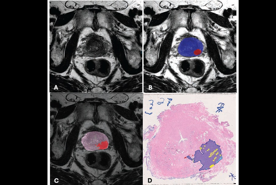
Gelikman and colleagues evaluated the impact of using a deep-learning AI model with bpMRI interpretations of prostate exams, tracking lesion- and patient-level clinically significant prostate cancer, cancer detection rates, and interreader agreement. Their study included six radiologist readers who interpreted bpMRI scans with and without AI assistance and 180 patients (120 in a case group, and 60 in a control group) who underwent bpMRI and prostate biopsy or radical prostatectomy between January 2013 and December 2022.
The researchers used pathology after radical prostatectomy as the reference standard for case group patients and negative 12-core systematic biopsies for control patients. They assessed the following measures: lesion-level sensitivity, positive predictive value (PPV), patient-level area under the receiver operating curve (AUC) for clinically significant prostate cancer and any prostate cancer detection, and interreader agreement for lesion-level PI-RADS scores and lesion size measurements.
Overall, the investigators found that the use of AI for bpMRI interpretations improved lesion-level PPV and interreader agreement but showed comparable AUC values and slightly lower lesion-level sensitivity compared to non-AI-assisted interpretations.
|
Assessment of use of AI with bpMRI exam interpretation |
||
|
Measure |
Interpretation without AI assistance |
Interpretation with AI assistance |
|
Clinically significant prostate cancer |
||
| Lesion-level PPV |
67.2% |
77.2% |
| Lesion-level sensitivity |
48% |
44.4% |
| Patient-level AUC |
0.83 |
0.82 |
|
Any prostate cancer |
||
| Lesion-level PPV |
69.4% |
80.9% |
| Lesion-level sensitivity |
44.9% |
41.7% |
| Patient-level AUC |
0.83 |
0.83 |
|
Interreader agreement |
||
| Lesion-level PI-RADS scores |
κ = 0.336 |
κ = 0.748 |
| Patient-level PI-RADS scores |
κ = 0.507 |
κ = 0.704 |
In any case, more investigation into the use of AI with bpMRI for prostate imaging is needed, according to the team.
“Future research should focus on further optimizing AI algorithms to improve lesion-level sensitivity without compromising specificity, further integrating AI into clinical workflows to enhance patient outcomes,” Gelikman and colleagues concluded.
The complete study can be found here.