 BBC
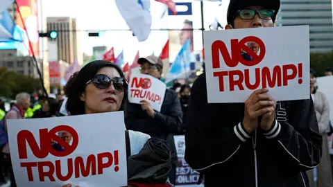
BBC“No Trump!” the rally of hundreds shouted, growing louder as it neared the United States embassy in the centre of South Korea’s capital Seoul.
A line of police buses stopped them…
 BBC
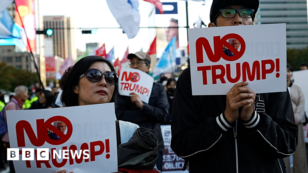
BBC“No Trump!” the rally of hundreds shouted, growing louder as it neared the United States embassy in the centre of South Korea’s capital Seoul.
A line of police buses stopped them…