Argentina’s National Council for Scientific and Technical Research, known as CONICET, has been pivotal in several of this year’s most thrilling scientific advancements. Several months back, they conducted a submarine expedition that turned…
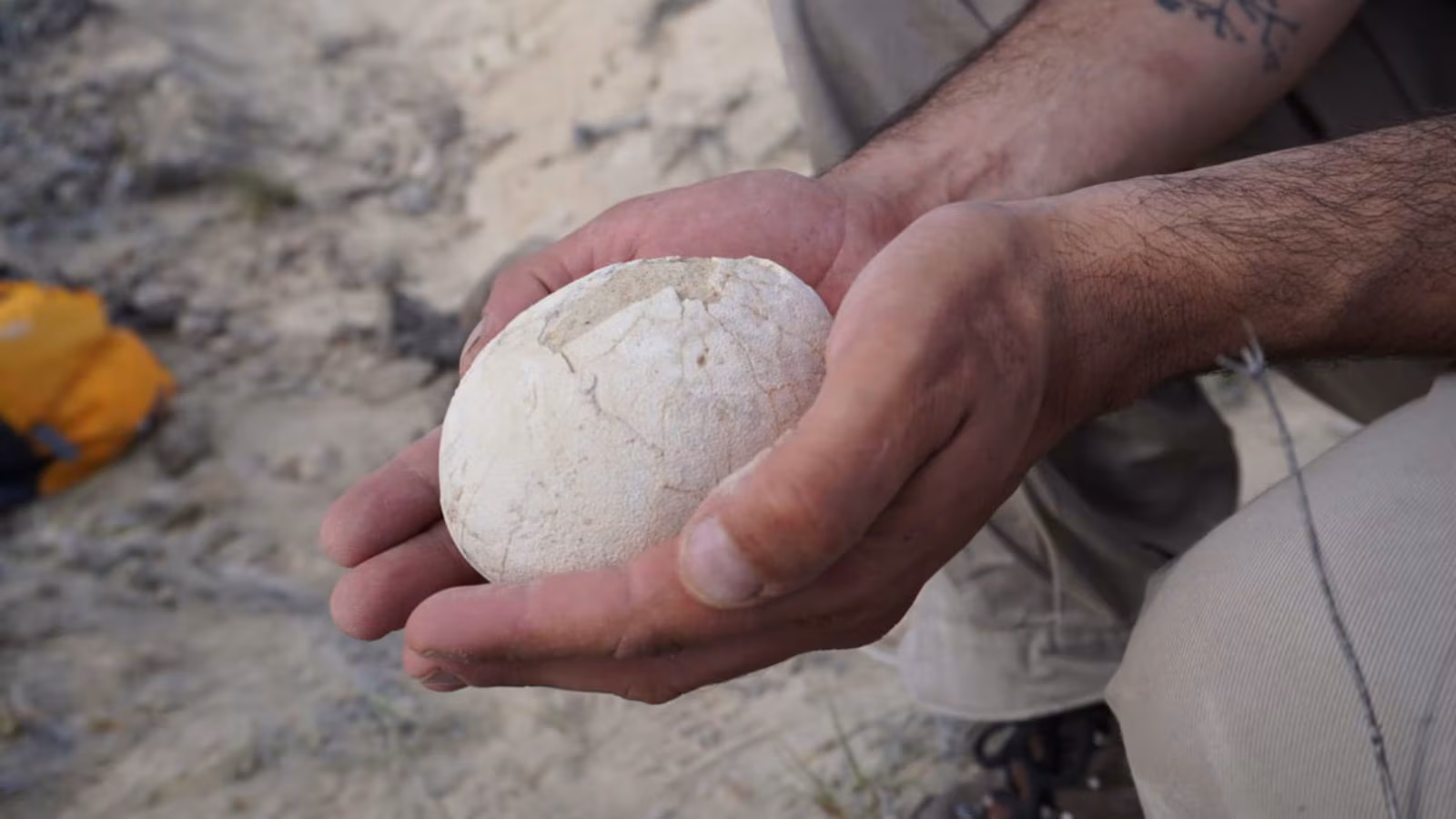
Pristine 70-million-year-old dinosaur egg unearthed in Patagonia; hints at ancient breeding ground | Technology News