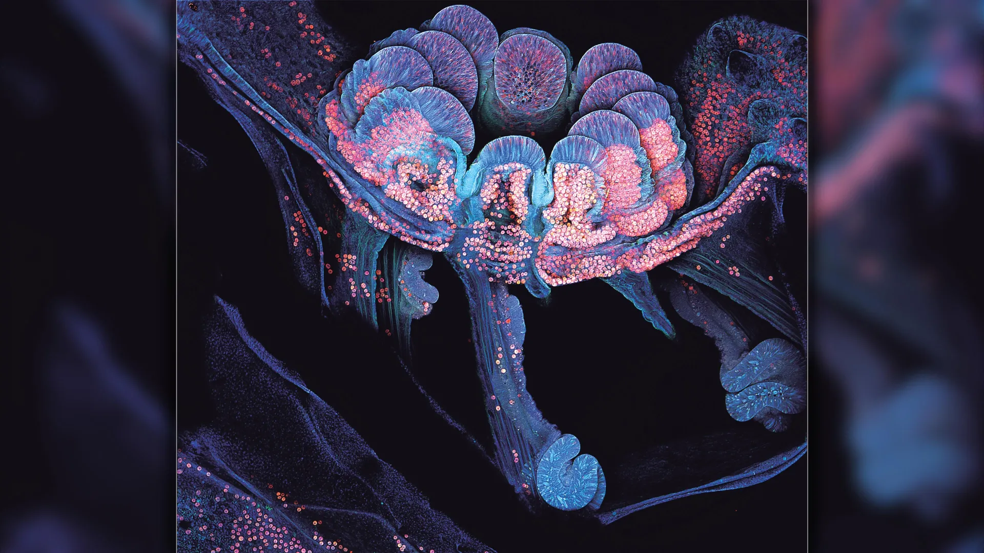
Bacteria strains and preparations
P. aeruginosa strains were isolated from a patient with chronic osteomyelitis at Peking University Third Hospital (Beijing, China) and identified by Genebang Technology Co., Ltd using genome sequencing. The bacterial strains were revived on Luria-Bertani (LB) agar and subsequently cultured at 37 °C overnight. The bacterial suspension was cultivated in LB broth at 37 °C for 4.5 h on a shaker at 200 rpm.
Isolation and enrichment of phages
Wastewater samples from the Third Hospital of Peking University were collected and centrifuged at 5,000 × g for 5 min at 4 °C in 2024. Phage isolation and purification were performed as described previously [9], with minor modifications. First, the centrifuged sewage and host bacteria culture were added to the LB liquid medium. The enrichment culture was subsequently incubated overnight at 37 °C, centrifuged (12,000 × g for 5 min), and the supernatant was filtered through a 0.22-µm sterile filter. The filtrate was serially diluted, mixed with host strains in molten semisolid soft agar (0.75% w/v agar), and poured onto solidified 1.5% w/v nutrient agar plates. After cooling and solidification, the plates were incubated overnight at 37℃ to assess plaque formation. The plaques were subsequently subjected to three rounds of plaque purification. The SM buffer (100 mM NaCl, 8 mM MgSO4.7H2O, and 50 mM Tris-HCl at pH 7.5) was used to store the purified phages and the double-layer agar (DLA) plaque assay was used to determine the phage titer [10].
Morphological characterization and phage potency determination
Phage morphology was characterized by transmission electron microscopy (TEM). Briefly, 15ul of the sample suspension was dropped onto the electron microscope mesh (AZH200; CHN) for 10 min of adsorption. Then, 20ul of uranyl acetate staining solution (22400, EMS) was added to the electron microscope mesh for 5 min. The excess solution was removed, and the grid was air-dried under an incandescent lamp. The images were captured by a TEM (JEM1400, jeol) at 80 kV.
A specific volume of the phage purification solution was serially diluted to 10− 1-10− 9 and was mixed with the bacterial solution in the logarithmic growth phase. The DLA method was used to culture the mixed solution overnight. Subsequently, the number of plaques on each plate was counted, and the phage titer was determined.
Determination of multiplicity of infection
The overnight-cultured host bacterial solution was transferred to 10 mL of LB medium (1:100 dilution) and cultured until reaching the logarithmic phase. Phage dilutions were mixed with an equal volume of host bacterial solution at different multiplicity of infection (MOI) values (10, 1, 0.1, 0.01, and 0.001). The mixture was transferred to sterilized LB liquid culture medium and incubated at 37 ℃ for 4 h. Subsequently, the mixture was centrifuged at 5,000 × g for 2 min. Afterward, the supernatant was collected. As described above, the phage titers in the supernatant for each MOI were determined, and the MOI yielding the highest phage titer was identified as the optimal MOI. The experiment was repeated in triplicate, and the mean value was calculated.
Temperature and PH stability
The temperature stability of the phage was assessed as follows. 1 mL each of phages (107 PFU/mL) solution was incubated in a water bath at 37 °C, 40 °C, 50 °C, 60 °C, and 70 °C. After 1 h of incubation, the potency was measured on a double-layer plate. The above experiment was repeated in triplicate.
To detect the stability of the phage in acidic and alkaline conditions, phages suspension at concentration of 105 PFU/mL were added to buffer solutions with pH values of 1.0, 2.0, 3.0, 4.0, 5.0, 6.0, 7.0, 8.0, 9.0, 10.0, 11.0, and 12.0, respectively. After incubation in a 37 °C water bath for 1 h, the phage titer was determined using the DLA method.
The phage titers at 37 °C (for temperature stability) and pH 7.0 (for pH stability) were used as the reference baselines for all comparisons. Furthermore, the control titers were determined to be 3 × 10⁷CFU/mL at 37 °C and 2 × 10⁵ CFU/mL at pH 7.0 under standard experimental conditions.
Adsorption curve
The phages at the optimal MOI were mixed with bacterial suspensions. Subsequently, the mixture was incubated on a shaker at room temperature (25 °C). Aliquots of 100 µL were collected from the mixture at 3-minute intervals and immediately centrifuged for 30 s. The supernatant was serially diluted, and the phage titer was determined using the DLA method. The test was repeated in triplicate.
One-step growth curve
P. aeruginosa cells were harvested by centrifugation, resuspended in fresh medium, and mixed with phages at the MOI of 10. The mixture was incubated on a shaker at 37 °C for 15 min. Aliquots of 100 µL were collected from the mixture at 10-minute intervals, and centrifuged (60s, 13,000×g). After removing the supernatant, the precipitate was resuspended in a liquid LB medium and centrifuged again to wash away any unabsorbed phages. 10 µL of the supernatant was rapidly serially diluted for phage titer determination using the DLA method. The phage titer (PFU/mL) was plotted against infection time (t) on the horizontal axis. A one-step growth curve was plotted on the y-axis to determine the incubation and lysis periods of the bacteriophage, and the burst size of each cell was calculated. The burst size was calculated using the following formula: Burst size = (number of phages at the highest point) / (total number of infected bacteria in the system).
Early biofilm formation and clearance assay
The crystal violet staining was used to detect the ability of bacteriophages to inhibit early biofilm formation [11]. After overnight incubation, the Pseudomonas aeruginosa bacterial solution was diluted 1:100, and 10 µL was added to each well of a 96-well plate along with 190 µL of TSB medium. Subsequently, 10 µL of bacteriophage (MOI = 1 or MOI = 0.1) was added to the experimental groups, followed by continuous cultivation at 37 °C for 72 h. After incubation, the medium was removed, and the wells were gently washed three times with PBS to remove non-adherent bacteria. The appropriate amount of methanol was added to each well plate and fixed for 30 min. After removing the methanol, the plates were air-dried for 30 min at 37 °C. An appropriate amount of 1% crystal violet was added to each well for 10 min at room temperature. Thereafter, 200 µ L of 33% acetic acid was added to each well to dissolve the crystal violet. Finally, the absorbance at 600 nm was measured using a microplate reader.
Whole genome sequencing and analysis
Phage DNA was extracted and its genome was sequenced using the IDNBSEQ-T7 (Illumina, San Diego, CA, USA) at Sciensano (Brussels, Belgium). To ensure the reliability of subsequent information analysis, the clean-read sequencing libraries were prepared with fastp, according to the manufacturer’s instructions. Trimming of the short reads was performed with fastp [12]. Low-quality reads and those with a high proportion of ambiguous bases (N) were discarded. De novo assembling was performed using metaSPAdes (version 3.1) [13]. Then, the clean reads were compared to assembled genome sequences to count the coverage rate using the BWA software. The Prokka was used to annotate the coding genes and tRNA of the genome [14]. Subsequently, the protein sequences were aligned against the NR database using BLASTp to identify homologous sequences. The functions of the open reading frames (ORFs) were analyzed using eggNOG-mapper software [15]. The genome map was generated using CGView (version 1.0).
To roughly estimate the relationships between related sequences and Pseudomonas phage PUTH1, the circular and rectangular proteomic tree was constructed by VIPtree (version 4.0). The analysis was reference dsDNA prokaryotic viruses [16]. Fifteen phages with the highest ViPTree tBLASTx genomic similarity (SG) scores were selected to construct a detailed rectangular proteomic tree. Furthermore, genome-genome comparison was conducted for PhiCHU with highest SG scores and phage PaP3 with second SG scores using ViPTree. Moreover, the gene sequences were aligned against the Virulence Factor Database (VFDB) with a stringent E-value cutoff of ≤ 1 × 10− 5 to predict potential virulence genes [17]. Antibiotic resistance genes were screened using ResFinder, applying thresholds of ≥ 60% coverage and ≥ 50% identity [18]. The genome sequence has been deposited in GenBank under the accession number: PV521973.
Statistical analysis
All statistical analyses were performed using GraphPad Prism 10 (GraphPad Software Inc., San Diego, CA, USA). Continuous variables were compared using one-way analysis of variance (ANOVA) between the three experimental groups. Data are presented as mean ± standard deviation (SD) from three independent biological replicates. Statistical significance was defined as *p < 0.05 and **p < 0.01.